Learning anatomy/Printable version
| This is the print version of Learning anatomy You won't see this message or any elements not part of the book's content when you print or preview this page. |
The current, editable version of this book is available in Wikibooks, the open-content textbooks collection, at
https://en.wikibooks.org/wiki/Learning_anatomy
Learning anatomy crew
Authors
[edit | edit source]We make it possible for people to read. Soon, your brain will be full of anatomy. User:Gffsgeys uses Grammarly to improve my mitsakes | mistakes. Red means that the word is spelled incorrectly. Green means that the word is spelled correctly.
Guidelines
[edit | edit source]1. At the moment the book is not about human body science experiments like the lungs in an empty water bottle.
2. We use photos as long as the photo shows the anatomy of the same organ but not videos (but these are only guidelines, so you can if you want).
3. Now learn about the editing rules.
4. Never add periods to a gallery.
5. Don’t do Bold text, Italic text, or Underline text unless you really must.
Now let’s get ready to make the book but please think about the guidelines.
By Gffsgeys (discuss • contribs) 01:35, 6 September 2021 (UTC) _____________________________________________________________________
User:Gffsgeys
[edit | edit source]_______________________________________________________________________
Books by Gffgeys
[edit | edit source]Templates by Samuel
[edit | edit source]Project pages by Samuel
[edit | edit source]
User:Xania
[edit | edit source]
| This user is a member of the Wikimedia OTRS team. (verify) |
| This user contributes using Opera. |
| This user is a recent changes patroller |
| …in. | Ending a sentence with a preposition is something this user is okay with. |
| This user is a member of the Counter-Vandalism Unit. |
|}
X8
|
A1
|
N1
|
I1
|
A1
|
Hi, I'm Xania. I've been here at Wikibooks since 2005 and presently I'm an administrator (since 2006) and check user (since 2014) here so happy to help with whatever problems you may have.
My specialties are linguistics (Romance, Slavic and Celtic languages and Basque), history, European geography, culture and politics.
I help out here with removing vandalism and spam, checking books for grammar or spelling mistakes, categorizing books, improving Cookbook pages by adding links and metric units and working on several books which I created or have contributed to.
Some Wikibooks that I am currently working on:
[edit | edit source]- English - Principal English book
- English for B2 Students - This book is intended for Upper Intermediate students.
- English in use - This book was previously known as "English" and is a Wikibook resource for English grammar and vocabulary.
- English_as_an_Additional_Language - This is an older book for those learning English as a Foreign Language.
- FCE English (Book I created but forgot about)
- Polish (Basic Polish Language Course)
- Italian (reviewing lessons, adding extra information)
- Basque (TBC)
- Japanese (TBC)
- Manx (TBC)
- Wikijunior Languages (inter-linking WJ Languages, WJ Europe and WJ South America projects)
- Wikijunior Languages/Manx Gaelic (Junior Guide to the Isle of Man's Language)
- Wikijunior Europe (Completed junior Guide to Europe) (Featured Book)
- Wikijunior World Religions
- Wikijunior World Heritage Sites (new book I'm developing for WJ about UNESCO heritage sites) create
- Wikijunior:Countries A-Z (adding more pictures)
- Wikijunior:Solar System (TBC)
- Wikijunior:South America (creating pages, updating info)
- Wikijunior:Fruit Alphabet (I'm adopting this abandoned book and trying to expand it)
- Wikijunior:Africa (a book I plan to develop further)
- High School Earth Science (Changing settings to only show reviewed page version, currently done units 1-4 and sub pages)
- Cookbook (Adding Metric Measurements to all Recipes)
- User:Xania/Sandbox (Recipes still without Metric Measurements)
- User:Xania/Sandbox2
My Activity on Other Wikis:
[edit | edit source]I am most active on Wikibooks but you may also find me on these Wiki projects (especially Wikipedia and Wikivoyage English sites and Wikipedia Italian site):
Wikibooks projects:
[edit | edit source]- English Wikibooks (home wiki & sysop)
Wikipedia projects:
[edit | edit source]Wiktionary projects:
[edit | edit source]Other Wikimedia projects:
[edit | edit source]Languages
[edit | edit source]I am an English language professional with experience teaching the language to native speakers and non-native speakers as well as developing materials for use by other teachers. I speak Italian to a reasonable degree. I also have a more limited knowledge of Russian, French and Polish.
My Basic 'Policies' on Wikibooks
[edit | edit source]I spend a lot of time fighting vandalism on Wikibooks. I am always willing to give people the benefit of the doubt, but I have no tolerance for malicious vandalism.
Contact me
[edit | edit source]You can often find me on IRC using the username Xania:
- #wikibooksconnect
- Alternatively leave me a message on my talk page and I'll get back to you as soon as I can.
Useful Links[edit | edit source]These are mainly a shortcut for pages that I commonly visit but which take forever to find because of the poor search feature on Wikibooks.
|
Useful Templates[edit | edit source]
Edit Warnings[edit | edit source]
|
User:Mbrickn
[edit | edit source]Intro
[edit | edit source]Howdy!
I mainly edit on:
- Wikibooks
- Wikivoyage
- Wikimedia Commons
- Wikidata
I occasionally also use:
- Wikipedia
- Wikiversity
Here I'm mostly interested in updating and modernizing older content. I'm also interested in improving technology and culture articles.
My Workspace
[edit | edit source]Most of the stuff here isn't really relevant to most people, aside from User:Mbrickn/Wikibooks tips, and is basically my Wikibooks scratchpad. If you're interested in what I'm working on though, feel free to peruse.
To do
[edit | edit source]- At some point I'd like to try a recipe for globi used at Saturnalia.
- Tyrokafteri dip
- Fix weird issues with Chip's Challenge/Printable version
- Split FLOSS Concept Booklet
- Fix printable version in Mega Man (classic series)
- Lunar Lander PDP demo packin and Easter egg for history of video games Wikibook
- Make strategy guide standardized box with logo, skill level, intended audience, etc. Not quite like infobox, but something similar.
User Pages
[edit | edit source]Work
[edit | edit source]- User:Mbrickn/Development
- User:Mbrickn/Book ideas
- User:Mbrickn/Strategy Guide Writing/
- User:Mbrickn/Wikibooks Oddities
- User:Mbrickn/Wikibooks Philosophy
Tracking
[edit | edit source]Draft
[edit | edit source]Misc
[edit | edit source]Wikibooks
[edit | edit source]This is my WikiBooks user page. Other sites may copy contents from 𝒲𝒾𝓀𝒾𝒷ℴℴ𝓀𝓈, but I am almost certainly not affiliated with them. I have a template at the top of this section that explains it nicer, but in case that template is broken on the mirror, this footnote text should remain.
User:Minorax
[edit | edit source]
Table version
This is the Table version of Learning anatomy.
Esophagus
 |
This is in the gastrointestinal anatomy chapter. |  |
Anatomy
[edit | edit source]The esophagus is a tube connecting the throat to the stomach. The parts of the esophagus are the upper esophagus, middle esophagus, and lower esophagus. The esophagus also passes through a hole in the diaphragm called the esophageal hiatus.


Function
[edit | edit source]The function of the esophagus is to move the swallowed food from the throat into the stomach. The lower esophageal sphincter or cardiac sphincter is between the lower esophagus and cardiac region of the stomach. Peristalsis happens throughout the gastrointestinal tract.
Stomach
|
This is in the gastrointestinal anatomy chapter. | |
Anatomy
[edit | edit source]The stomach is a J-shaped ”bag” that connects the esophagus to the small bowel. The parts of the stomach are the cardiac region, gastric fundus, gastric body, and pyloric region. The pyloric region is made up of the pyloric antrum, and the pyloric canal.

Function
[edit | edit source]The first sphincter of the stomach is the cardiac sphincter or lower esophageal sphincter which guards the entrance to the stomach. If the sphincter weakens, stomach contents can damage the lining of the esophagus and cause acid reflux so, please take care of your lower esophageal sphincter.
The stomach has folds called rugae which look wrinkled when no food is in the stomach. The stomach stores food. If the food is poisonous, it triggers the vomiting reflex. In this case, the body protects itself from the acid. But if it’s not poisonous, the food continues to go. The stomach will make acid, digestive enzymes, and mucin. Hydrochloric acid breaks down all foods into chyme. Amylase breaks down starch. Pepsinogen is the inactive form of pepsin. When pepsinogen reaches the acidic ph of the stomach, it turns into pepsin which digests proteins. Gastric lipase breaks down lipids. Hydrochloric acid in the stomach kills germs. Gastrin and somatostatin are hormones. Gastrin inhibits acid secretion. Somatostatin does a lot of things.
Small bowel
Stomach - Large bowel Template:Toc
Anatomy
[edit | edit source]The small bowel or small intestine connects the stomach to the large intestine. The parts of the small intestine are the duodenum, jejunum, and ileum. 
Function
[edit | edit source]The small intestine absorbs most of the nutrients from the chyme. The pyloric sphincter lets only a tiny bit of chyme go into the small intestine at a time. The small finger-like projections are called villi. Villi help in the absorption. The jejunum absorbs sugars, amino acids, and fatty acids. The ileum absorbs bile acids, fluid, and vitamin B12.
Large bowel
Anatomy
[edit | edit source]The large bowel or large intestine is made up of the vermiform appendix, the cecum, and the colon. The colon is made up of the ascending colon, transverse colon, descending colon, and sigmoid colon. The large intestine connects the small intestine to the rectum.

Function
[edit | edit source]The cecum absorbs fluids and salts. The vermiform appendix is a storage for the gut bacteria that help with digestion. Gut bacteria is an example of a good bacteria. The colon absorbs water and electrolytes.
Rectum and anus
Anatomy
[edit | edit source]The rectum and anus connect the large intestine to the external body. There is also an anal canal that goes between them.

Defecation
[edit | edit source]The function of the rectum and anus is defecation. In this section, you will learn about the defecation reflex. The rectum stores the feces that the colon made. The nerve signals then tell the brain that you need to go into the bathroom. Then the feces go into the anus. There are 2 sphincters. The first is the internal sphincter and we can't control it. The last one is the external sphincter and we control it. Babys can't control their external sphincter so they do it in their diapers.
Liver
Accessory organs
_______________________________________________________________________
Rectum and anus - Biliary tract
Anatomy
[edit | edit source]The liver is made up of the left lobe, right lobe, caudate lobe, and quadrate lobe. The ligaments are the falciform ligament and the coronary ligament. They are not real ligaments. Instead, they are a piece of peritoneal membrane holding the liver. The lobes of the liver are divided into 8 segments.


Function
[edit | edit source]The liver has many functions and is a critical part of the digestive system. Did you know that the liver has more than 500 functions and can do 200 functions at the same time!? The liver makes bile, stores nutrients and performs metabolism. Bile is made by hepatocytes which are also called liver cells.
Biliary tract
Accessory organs
_______________________________________________________________________
Anatomy
[edit | edit source]The biliary tract is made up of the liver, gallbladder, and bile ducts.

Function
[edit | edit source]The hepatocytes make bile. After that, the bile goes into the bile canaliculi and then the bile goes into the interlobular ducts and then the bile goes into the intrahepatic ducts and then the bile moves into the right and left hepatic ducts and then the bile moves into the common hepatic duct and then the bile goes into the cystic duct and then the bile goes into the gallbladder. The gallbladder stores bile until needed. When needed, cholecystokinin triggers the bile to go out of the gallbladder and then into the common bile duct. Then the bile moves into the duodenum where the bile emulsifies fat.
Pancreas exocrine
Accessory organs
_______________________________________________________________________
Biliary tract - Pancreas endocrine
Anatomy
[edit | edit source]Acinar cells in the pancreas.

Function
[edit | edit source]Acinar cells make digestive enzymes and bicarbonate. Bicarbonate neutralizes the hydrochloric acid from the stomach. Amylase breaks down starch. Lipase breaks down lipids. Trypsin breaks down proteins. Chymotrypsinogen is activated by trypsin to make chymotrypsin. Chymotrypsin breaks proteins in the chyme.
Pancreas endocrine
Accessory organs
_______________________________________________________________________
Anatomy
[edit | edit source]Endocrine cells in the pancreas.
Function
[edit | edit source]Beta cells make insulin. Alpha cells make glucagon. Insulin is like a key for opening up the cells so that glucagon can get in.
Spleen
Anatomy
[edit | edit source]The red and white pulps.

Function
[edit | edit source]The spleen makes bilirubin which is sent to the liver to make bile. The spleen also fights off sickness.
Heart
Cardiovascular system
_______________________________________________________________________
Anatomy
[edit | edit source]The heart is inside the chest. The heart consists of the right atrium, left atrium, right ventricle, and left ventricle.

Function
[edit | edit source]The heart pumps blood through the body. The blue deoxygenated blood moves through the right atrium and then into the right ventricle and then into the lungs. This is where it mixes with the oxygen and it turns the blood red. The red blood travels all over the body. Then the oxygen has been used so it travels back to the heart and the cycle repeats.
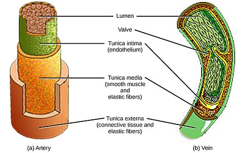
Artery
Cardiovascular system
_______________________________________________________________________
Anatomy
[edit | edit source]The arteries are made up of 3 layers which are the intima, media, and adventitia. The artery is made up of the endothelium, smooth muscle, and epithelium.

Function
[edit | edit source]The arteries move oxygenated blood away from the heart and to the entire body.
Arteriole
Cardiovascular system
_______________________________________________________________________
Anatomy
[edit | edit source]Just like the arteries, the arterioles have 3 layers called the adventitia, media, and intima.

Function
[edit | edit source]The arterioles move blood from the arteries to the capillaries.
Capillary
Cardiovascular system
_______________________________________________________________________
Anatomy
[edit | edit source]The capillaries are broken into 3 types which are the continuous, fenestrated, and sinusoidal capillaries.

Function
[edit | edit source]The capillaries move blood from the arterioles to the venules.
Vein
Cardiovascular system
_______________________________________________________________________
Anatomy
[edit | edit source]Just like the other blood vessels, the parts of the vein are the intima, media, and adventitia.

Function
[edit | edit source]Oxygen has been used so it travels back to the heart through the veins.
Venule
Cardiovascular system
_______________________________________________________________________
Anatomy
[edit | edit source]The venules are made up of the intima, media, and adventitia.
Function
[edit | edit source]The venules move blood from the capillaries to the veins.
Kidney
Accessory organs
_______________________________________________________________________
Anatomy
[edit | edit source]The kidneys are not just in kids. They are also in adults too. So why are they called kid
-ney? The answer is that the word kidney is probably a compound of the Old English cwið ”womb,” and ey, “egg,” describing the shape of the organ.

Function
[edit | edit source]The kidneys filter your blood and removes waste.
Illustrations
[edit | edit source]-
 Diagram of the Kidneys

Nephron
Anatomy
[edit | edit source]The parts of the nephron are the bowman’s capsule, proximal tubule, a loop of Henle, distal tubule, and collecting tube.

Function
[edit | edit source]The urine starts in the bowman's capsule and then into the proximal tubule and then into the loop of Henle and then into the distal tubule and then into the collecting tubes which takes the urine to the ureters.
Ureter
Anatomy
[edit | edit source]The parts of the ureter are the abdominal ureter, pelvic ureter, and intravesical or intramural ureter.
.jpg)
Function
[edit | edit source]The ureters carry urine from the kidneys to the bladder.
Bladder
Anatomy
[edit | edit source]The bladder or urinary bladder is made up of the dome or apex, neck, body, and fundus. The bladder is between the ureter and urethra.

Function
[edit | edit source]The detrusor muscle expands when filled with urine. Then the nerve signals tell the brain that you need to run to the bathroom. When emptying, the detrusor muscle squeezes the urine to the urethra. Check out the urethra page to learn more. Make sure to read the kidney, nephron, and ureter pages too.
Urethra
Anatomy
[edit | edit source]Male and female urethras are different.
Male
[edit | edit source]The first thing at the bottom of the urethra is called the penis. Then there is the spongy urethra and then there is the membranous urethra. The prostatic urethra is last.
Female
[edit | edit source]The parts of the female urethra are the muscular, erectile, and mucous parts but no penis.
.png)
Function
[edit | edit source]The internal urethral sphincter opens itself. The external sphincter is what we control to open and when that sphincter is open it goes into the toilet. It passes through the penis in men.
Muscle
Anatomy
[edit | edit source]There are 650 muscles in the body made by myocytes.
Function
[edit | edit source]Skeletal muscles control movement so we can walk, run, and jump and even breathing and chewing needs muscles. Smooth muscle is what we can’t control. Cardiac muscle is found only in the heart.
Illustrations
[edit | edit source]-
 Skeletal Muscle diagram
 -
 Smooth Muscle diagram
 -
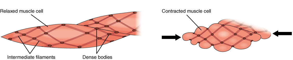 Smooth Muscle contraction diagram
 -
 Cardiac Muscle diagram


Bone
Anatomy
[edit | edit source]The parts of the bone are the periosteum, compact bone, and spongy bone.

Function
[edit | edit source]Bones help to support muscles, protect organs, and even hold your teeth. Bones help so that we wouldn't be 1 flat puddle on the ground.
Ear
Anatomy
[edit | edit source]The parts of the ear are:
- Pinna
- Ear canal
- Eardrum
- Ossicles
- Hammer
- Anvil
- Stirrup
- Cochlea

Function
[edit | edit source]When sound waves are made the pinna (auricle) gathers them up and then the sound waves move into the ear canal (external auditory meatus, EAM). The sound waves then vibrate the eardrum (tympanic membrane) which strikes the hammer (malleus) bone which moves the anvil (incus) bone and the stirrup (stapes) bone. The stapes vibrates the liquid that fills the cochlea. The hair cells at the bottom detect the highest sounds. The hair cells in the middle detect lower sounds. The hair cells at the top control the lowest sounds. This gets going to the auditory nerve and then into the brain where you hear the sound.
Illustrations
[edit | edit source]-
 the pinna of the ear
 -
 ear canal highlighted
 -
 tympanic membrane
 -
 ossicles
 -
 cochlea


Nose
Anatomy
[edit | edit source]The parts of the nose are:
- Nasion
- Root
- Bridge
- Ala
- Apex
- Nostril
- Columella
- Nasal cavity
- Vestibule
- Olfactory region
- Respiratory region
Function
[edit | edit source]The air goes inside the nasal cavity. The nasal cavity warms the air and traps dust particles with tiny hairs and mucous. The smell of things in the air is caused by the olfactory tract which sends signals to the brain about the smell.
Mouth
Tooth
[edit | edit source]Anatomy
[edit | edit source]The teeth (singular: tooth) are made up of parts shown below.
- Crown
- Enamel
- Dentin
- Neck
- Gingiva
- Pulp cavity
- Pulp
- Root
- Cementum
- Root canal
- Periodontal ligament
- Nerves and blood vessels
- Alveolar bone
- Jaw
- Maxillae
- Mandible

Function
[edit | edit source]The teeth function to chew food. The types of teeth are the incisors, canines, premolars, and molars. Incisors cut the food. The canines grip the food. The premolars and molars (from Latin: mola = grindstone) grind the food. A baby has 20 teeth, but they are not visible until they erupt through the gingiva (gums) and an adult has 32 teeth. Maxillary (upper) teeth and their associated periodontal ligament are innervated by the superior alveolar nerves, branches of the maxillary division, termed the posterior superior alveolar nerve, anterior superior alveolar nerve, and the variably present middle superior alveolar nerve. The teeth help us speak.
Salivary gland
[edit | edit source]Anatomy
[edit | edit source]The parotid, sublingual, and submandibular glands are major salivary glands. The other ones are minor salivary glands.
Function
[edit | edit source]The salivary glands make saliva. Saliva travels through ducts and then into the mouth.
Saliva
[edit | edit source]Saliva has alpha-amylase which breaks down starch and lingual lipase which breaks lipids. Saliva also cleans the mouth.
Lingua
[edit | edit source]Anatomy
[edit | edit source]The lingua (tongue) is made up of the base (root), body, and apex.
Function
[edit | edit source]The lingua helps in speech. Saliva helps taste the food. The chemicals from the food travel from the lingual papillae to the taste buds. The lingua and saliva from the food bolus. It gets sent down the pharynx.
Air
[edit | edit source]The mouth is also a passage for air.
Throat
Pharynx
[edit | edit source]Anatomy
[edit | edit source]The pharynx is made up of the nasopharynx, oropharynx, and laryngopharynx.

Function
[edit | edit source]The pharynx brings food from the oral cavity to the esophagus. The pharynx also brings air from the nasal and oral cavities to the larynx.
Larynx
[edit | edit source]Anatomy
[edit | edit source]The parts of the larynx are the epiglottis, thyroid, cricoid, arytenoids, cuniform, corniculates, infrahyoid, suprahyoid, thyrohyoid membrane, lateral thyrohyoid ligament, quadrangular membrane, cricothyroid ligament complex, and posterior cricoarytenoid ligament.
Function
[edit | edit source]The larynx helps in speech. The larynx moves air from the pharynx to the trachea. The epiglottis prevents food from entering the airway.
Epidermis
Anatomy
[edit | edit source]
The parts of the epidermis are:
- Stratum basale
- Stratum spinosum
- Stratum granulosum
- Stratum lucidum
- Stratum corneum
Function
[edit | edit source]The epidermis prevents microbes from entering the body. The epidermis is also waterproof, preventing the ingress of liquids which would otherwise wreck havoc on the chemistry body. After all, how would you wash your hands if they were not waterproof?
Dermis
Anatomy
[edit | edit source]The dermis is made up of the papillary dermis and the reticular dermis. It consists of collagen, elastin, blood capillaries, lymph vessels, sweat glands, sebaceous glands, nerve endings, and hair follicles.
Function
[edit | edit source]The dermis makes sweat and regulates the body’s temperature, it produces oil, it makes hair grow, feels things and if it is too hot or too cold, your body tells you to get away from it and the dermis also protects the body from harmful pathogens, keeping the skin in shape, and distributing blood so that it can feed the skin, removes toxins, and supplies the epidermis with blood.
Test
Quiz
[edit | edit source]

You have random organs. You have to type the name. You also have to describe if the the the organ is needed to live. Your score will be on Wednesday. Answers will be cleared 10 days after being scored so other people can answer. Please edit visually. Please put your user name so that I know who did it. First, I will read your user page and then give you a score. I will remind you that you need to put your user name if you didn't. I will also put it on your talk page in case you forget.
Facts
[edit | edit source]