Cosmetic Science/Printable version
| This is the print version of Cosmetic Science You won't see this message or any elements not part of the book's content when you print or preview this page. |
The current, editable version of this book is available in Wikibooks, the open-content textbooks collection, at
https://en.wikibooks.org/wiki/Cosmetic_Science
Welcome
Welcome
[edit | edit source]This book is intended to be a comprehensive teaching tool for newcomers to cosmetic science. In addition, it also serves as a convenient reference for gurus and formulators. For beginners, the best approach is to read the book in order. Check out the Ingredients Glossary when you need formulating tips for a particular ingredient.
Guidelines for Contributors
[edit | edit source]- Cite all sources and use reputable references (See Introduction: Research Methods).
- Explain concepts in simplest terms, but include technical terms with an explanation.
- Use the Discussion tab to raise issues about accuracy, organization, new topics, etc.
- Add yourself to the list of contributors.
Purpose
Why Learn Cosmetic Science?
[edit | edit source]The cosmetics industry is swamped with marketing hype, often promoting ingredients or treatments with little to no evidence of efficacy. The internet is filled with claims about home remedies and industrial ingredients, but those claims tend to have just as little evidence. With known scientific principles and the available scientific literature online, we can predict what will work without relying on unbacked claims. Here's how learning cosmetic science can change your life as both a shopper and DIYer.
- Get Better Results Listening to testimonials, marketing labels like "Dermatologist Tested", and advertisements are ineffective ways to predict how well a product would work for you. A product's efficacy is actually based on a few variables: the active ingredients, concentration, and your personal skin attributes. Too often, people confuse active ingredients with "claims ingredients". Claims ingredients are ingredients that provide no functional benefit to the user, but are added to the product in order to impress consumers[1]. Knowing how to distinguish the effective ingredients and how much of it is suitable for your body makes it much easier to find what works for you. Besides shopping, cosmetic science allows you to be a better home formulator. To make a quality product, you need to know about ingredient compatibility, the causes of skin irritation, how to create the desired texture, and so on. All of this is based on the underlying chemical properties of the ingredients and their biochemical interactions with the body.
- Save Time and Money By looking for the right ingredients and concentration, you do not have to spend as much time experimenting with various products. Knowing why a particular product works well means that you don't have to be loyal to a particular brand and you have more flexibility in where you shop. In addition, it is often more cost effective to make cosmetics at home, especially if you want to use products over your entire body. Hopefully, this wikibook can help you further save time and money by preventing formulating mistakes and needless experimentation.
- Avoid Pseudoscientific Thinking There are many myths about cosmetics perpetuated both because of ignorance and a profit motive. These myths often prey on fear of "chemicals", "toxins", the "unnatural", and anything else unknown. This wikibook will not focus on debunking these myths. However, it will provide a basic understanding of how various ingredients can affect the body, as well as explain the federal regulations in concerns to cosmetics. This knowledge plus the resources listed in Introduction: Research Methods should allow you to assess the true risks of cosmetic ingredients.
Scientific Outreach
[edit | edit source]Cosmetic science has great potential for engaging more people, especially young women, in STEM topics. It encompasses both biology and chemistry, and relates these subjects to very common concerns. In the US, the cosmetics industry has an annual revenue of about $57 billion with the following market share:[2]
- Skincare 34.1 %
- Haircare 24.1 %
- Make-Up 16.6 %
- Perfumes 12.7 %
- Toiletries, deodorants 11.2 %
- Oral Cosmetics 1.3 %
Additionally, 58% of girls between 8 and 18 wear make-up. Among these consumers, 65% of them started between the ages of 8 and 13 [2]. An even greater proportion of teenagers probably use skincare, considering the market share size in comparison and the prominence of marketing for acne-related products. This makes cosmetics a highly common interest amongst a group whom educators are trying to imbue with appreciation and understanding of the sciences.
Through cosmetic science, people can learn to experiment whether by formulating or testing products. They can become more scientifically literate by learning how to read scientific studies to address their skin concerns. They can become better critical thinkers by looking beyond marketing claims and using the back label to judge the potential efficacy of the product. Cosmetic science could be one step towards more people realizing that science is not restricted to professionals in lab coats. It is a discipline that can be applied to many aspects of daily life.
Scope of this book
[edit | edit source]After reading this book readers should have a full understanding of how cosmetics work and how to formulate products at home. This book aims to explain all related concepts at the level of high school chemistry and biology. However, there will be a brief explanation of organic chemistry concepts.
Not included in this book:
- Thorough evaluations of ingredient safety and efficacy (See links at Introduction: Research Methods for more information)
- Specific formulas to follow, other than occasional examples
- Product recommendations
References
[edit | edit source]- ↑ [1], Romanowski, P. (n.d.). 7 Types of Cosmetic Story Ingredients. Retrieved June 25, 2017, from http://chemistscorner.com/7-types-of-cosmetic-story-ingredients/
- ↑ a b [2], "Cosmetic Industry Statistics." Statistic Brain. February 05, 2016. Accessed June 21, 2017. http://www.statisticbrain.com/cosmetic-industry-statistics/.
Research Methods
Databases
[edit | edit source]Databases are a great place to learn more about any particular subject. They compile information from various sources into one location, and provide search tools to aid research. All of the databases listed here are open-access and free to the public. In addition, the databases here are credible sources. Credible means that the databases:
- Use neutral language
- Are supported by scientific or governmental institutions
- Use non-subjective, clearly defined metrics
- Have well-documented references
Chemical Properties
[edit | edit source]Ingredients Usage and Safety
[edit | edit source]- Cosmetic Ingredients Review
- Cosmetic Info
- CosIng (maintained by the EU)
Clinical Trials and Scientific Research
[edit | edit source]- The Journal of the American Medical Association (requires account registration)
- PubMed
- Semantic Scholar
Quality of Scientific Research
[edit | edit source]
...
Governmental Regulations
[edit | edit source]...
Professionals
[edit | edit source]In addition to these institutions, individuals working in the industry may share their insights through articles, books, or podcasts. However, the differences between education and job capacity of various professionals may make them unqualified to speak on the same topics. These experts have a lot of useful insights from working in their field. However when experts start speaking outside of their field, they should provide additional references to remain credible.
Cosmetic Chemists
[edit | edit source]- Typical Educational Attainment: Bachelors in Chemistry, Chemical Engineering, Biology, or Microbiology[1]
- Typical coursework: General Chemistry, Organic chemistry, Inorganic chemistry, Physical chemistry [2]
- Typical Job Roles: formulation, quality control, analytics, process engineering, raw material synthesis, regulation, and sales[1]
- Scope: cosmetic industry practices, formulation, safety concerns
Dermatologists
[edit | edit source]- Typical Educational Attainment: doctoral degree in medicine (MD) or osteopathic medicine (DO), 1 year internship, and 3 years in dermatology residency.[3]
- Typical Coursework: human anatomy and physiology, pharmacology, biochemistry, microbiology, pathology, genetics, ageing, immunology, nutrition [4]
- Typical Job Roles: surgical procedures, skin diagnosis, injections, hair transplants [3]
- Scope: skin and hair conditions, carcinogenic hazards, surgical procedures, fillers and botox,
Estheticians
[edit | edit source]- Typical Educational Attainment: state-issued license after completion of around 600 credit hours at a cosmetology school and passing state exam (except in Connecticut) [5]
- Typical Coursework: human anatomy and physiology, massage, skin histology, makeup application, exfoliation, cleansing , sanitation [5]
- Typical Job Roles: massage, body treatment, facial, and nail care [5]
- Scope: massage, exfoliation, product recommendations, makeup looks, spa treatments
References
[edit | edit source]- ↑ a b [3], Romanowski, P. (n.d.). How to Become a Cosmetic Chemist. Retrieved June 25, 2017, from http://chemistscorner.com/how-to-become-a-cosmetic-chemist/.
- ↑ [4], B.S. Chemistry. (n.d.). Retrieved June 25, 2017, from http://chemistry.berkeley.edu/ugrad/degrees/chem.
- ↑ a b [5]Aad.org. (2017). Why see a dermatologist | American Academy of Dermatology. [online] Available at: https://www.aad.org/public/diseases/why-see-a-dermatologist [Accessed 1 Jul. 2017]
- ↑ [6], Usmle.org. (2017). United States Medical Licensing Examination | Step 1. [online] Available at: http://www.usmle.org/step-1/#content-outlines [Accessed 1 Jul. 2017]
- ↑ a b c [7],Education, Certification & Licensing. (n.d.). Retrieved June 25, 2017, from http://howtobecomeanesthetician.com/
Skin Anatomy and Physiology
Epidermis
[edit | edit source]
...
Dermis
[edit | edit source]...
Subcutaneous Tissue
[edit | edit source]...
Skin Conditions
| This page was imported and needs to be de-wikified. Books should use wikilinks rather sparsely, and only to reference technical or esoteric terms that are critical to understanding the content. Most if not all wikilinks should simply be removed. Please remove {{dewikify}} after the page is dewikified. |

A skin condition, also known as cutaneous condition, is any medical condition that affects the integumentary system—the organ system that encloses the body and includes skin, hair, nails, and related muscle and glands.[1] The major function of this system is as a barrier against the external environment.[2]
Conditions of the human integumentary system constitute a broad spectrum of diseases, also known as dermatoses, as well as many nonpathologic states (like, in certain circumstances, melanonychia and racquet nails).[3][4] While only a small number of skin diseases account for most visits to the physician, thousands of skin conditions have been described.[5] Classification of these conditions often presents many nosological challenges, since underlying causes and pathogenetics are often not known.[6][7] Therefore, most current textbooks present a classification based on location (for example, conditions of the mucous membrane), morphology (chronic blistering conditions), cause (skin conditions resulting from physical factors), and so on.[8][9]
Clinically, the diagnosis of any particular skin condition is made by gathering pertinent information regarding the presenting skin lesion(s), including the location (such as arms, head, legs), symptoms (pruritus, pain), duration (acute or chronic), arrangement (solitary, generalized, annular, linear), morphology (macules, papules, vesicles), and color (red, blue, brown, black, white, yellow).[10] The diagnosis of many conditions often also requires a skin biopsy which yields histologic information[11][12] that can be correlated with the clinical presentation and any laboratory data.[13][14] The introduction of cutaneous ultrasound has allowed the detection of cutaneous tumors, inflammatory processes, nail disorders and hair diseases.[15]
Layer of skin involved
[edit | edit source]The skin weighs an average of 4 kg (8.8 lb), covers an area of 2 m2 (22 sq ft), and is made of three distinct layers: the epidermis, dermis, and subcutaneous tissue.[1] The two main types of human skin are glabrous skin, the non-hairy skin on the palms and soles (also referred to as the "palmoplantar" surfaces), and hair-bearing skin.[16] Within the latter type, hairs in structures called pilosebaceous units have a hair follicle, sebaceous gland, and associated arrector pili muscle.[17] In the embryo, the epidermis, hair, and glands are from the ectoderm, which is chemically influenced by the underlying mesoderm that forms the dermis and subcutaneous tissues.[18][19][20]
Epidermis
[edit | edit source]
The epidermis is the most superficial layer of skin, a squamous epithelium with several strata: the stratum corneum, stratum lucidum, stratum granulosum, stratum spinosum, and stratum basale.[21] Nourishment is provided to these layers via diffusion from the dermis, since the epidermis is without direct blood supply.[22] The epidermis contains four cell types: keratinocytes, melanocytes, Langerhans cells, and Merkel cells. Of these, keratinocytes are the major component, constituting roughly 95% of the epidermis.[16] This stratified squamous epithelium is maintained by cell division within the stratum basale, in which differentiating cells slowly displace outwards through the stratum spinosum to the stratum corneum, where cells are continually shed from the surface.[16] In normal skin, the rate of production equals the rate of loss; about two weeks are needed for a cell to migrate from the basal cell layer to the top of the granular cell layer, and an additional two weeks to cross the stratum corneum.[23]
Dermis
[edit | edit source]The dermis is the layer of skin between the epidermis and subcutaneous tissue, and comprises two sections, the papillary dermis and the reticular dermis.[24] The superficial papillary dermis interdigitates with the overlying rete ridges of the epidermis, between which the two layers interact through the basement membrane zone.[24] Structural components of the dermis are collagen, elastic fibers, and ground substance also called extra fibrillar matrix.[24] Within these components are the pilosebaceous units, arrector pili muscles, and the eccrine and apocrine glands.[21] The dermis contains two vascular networks that run parallel to the skin surface—one superficial and one deep plexus—which are connected by vertical communicating vessels.[21][25] The function of blood vessels within the dermis is fourfold: to supply nutrition, to regulate temperature, to modulate inflammation, and to participate in wound healing.[26][27]
Subcutaneous tissue
[edit | edit source]The subcutaneous tissue is a layer of fat between the dermis and underlying fascia.[5] This tissue may be further divided into two components, the actual fatty layer, or panniculus adiposus, and a deeper vestigial layer of muscle, the panniculus carnosus.[16] The main cellular component of this tissue is the adipocyte, or fat cell.[5] The structure of this tissue is composed of septal (i.e. linear strands) and lobular compartments, which differ in microscopic appearance.[21] Functionally, the subcutaneous fat insulates the body, absorbs trauma, and serves as a reserve energy source.[5]
Diseases of the skin
[edit | edit source]Diseases of the skin include skin infections and skin neoplasms (including skin cancer).[28]
History
[edit | edit source]In 1572, Geronimo Mercuriali of Forlì, Italy, completed De morbis cutaneis (translated "On the diseases of the skin"). It is considered the first scientific work dedicated to dermatology.
Diagnoses
[edit | edit source]The physical examination of the skin and its appendages, as well as the mucous membranes, forms the cornerstone of an accurate diagnosis of cutaneous conditions.[29] Most of these conditions present with cutaneous surface changes termed "lesions," which have more or less distinct characteristics.[30] Often proper examination will lead the physician to obtain appropriate historical information and/or laboratory tests that are able to confirm the diagnosis.[29] Upon examination, the important clinical observations are the (1) morphology, (2) configuration, and (3) distribution of the lesion(s).[29] With regard to morphology, the initial lesion that characterizes a condition is known as the "primary lesion," and identification of such a lesions is the most important aspect of the cutaneous examination.[30] Over time, these primary lesions may continue to develop or be modified by regression or trauma, producing "secondary lesions."[1] However, with that being stated, the lack of standardization of basic dermatologic terminology has been one of the principal barriers to successful communication among physicians in describing cutaneous findings.[21] Nevertheless, there are some commonly accepted terms used to describe the macroscopic morphology, configuration, and distribution of skin lesions, which are listed below.[30]
Lesions
[edit | edit source]Primary lesions
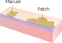
[edit | edit source]- Macule: A macule is a change in surface color, without elevation or depression and, therefore, nonpalpable, well or ill-defined,[31] variously sized, but generally considered less than either 5[31] or 10 mm in diameter at the widest point.[30]
- Patch: A patch is a large macule equal to or greater than either 5 or 10 mm across,[30] depending on one's definition of a macule.[1] Patches may have some subtle surface change, such as a fine scale or wrinkling, but although the consistency of the surface is changed, the lesion itself is not palpable.[29]
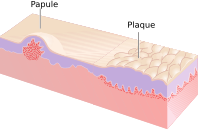
- Papule: A papule is a circumscribed, solid elevation of skin with no visible fluid, varying in size from a pinhead to less than either 5[31] or 10 mm in diameter at the widest point.[30]
- Plaque: A plaque has been described as a broad papule, or confluence of papules equal to or greater than 10 mm,[30] or alternatively as an elevated, plateau-like lesion that is greater in its diameter than in its depth.[29]
- Nodule: A nodule is morphologically similar to a papule in that it is also a palpable spherical lesion less than 10 mm in diameter. However, it is differentiated by being centered deeper in the dermis or subcutis.
- Tumour: Similar to a nodule but larger than 10 mm in diameter.
- Vesicle: A vesicle is small blister,[32] a circumscribed, fluid-containing, epidermal elevation generally considered less than either 5[31] or 10 mm in diameter at the widest point.[30] The fluid is clear serous fluid.
- Bulla: A bulla is a large blister,[32] a rounded or irregularly shaped blister containing serous or seropurulent fluid, equal to or greater than either 5[31] or 10 mm,[30] depending on one's definition of a vesicle.[1]
- Pustule: A pustule is a small elevation of the skin containing cloudy[29] or purulent material (pus) usually consisting of necrotic inflammatory cells.[30] These can be either white or red.
- Cyst: A cyst is an epithelial-lined cavity containing liquid, semi-solid, or solid material.[31]
-
Relative incidence of skin cysts
- Wheal: A wheal is a rounded or flat-topped, pale red papule or plaque that is characteristically evanescent, disappearing within 24 to 48 hours. The temporary raised bubble of taut skin on the site of a properly-delivered intradermal injection is also called a welt, with the ID injection process itself frequently referred to as simply "raising a wheal" in medical texts.[31]
- Telangiectasia: A telangiectasia represents an enlargement of superficial blood vessels to the point of being visible.[29]
- Burrow: A burrow appears as a slightly elevated, grayish, tortuous line in the skin, and is caused by burrowing organisms.[29][30]
Secondary lesions
[edit | edit source]- Scale: dry or greasy laminated masses of keratin[30] that represent thickened stratum corneum.[29]
- Crust: dried sebum, pus, or blood usually mixed with epithelial and sometimes bacterial debris.[31]
- Lichenification: epidermal thickening characterized by visible and palpable thickening of the skin with accentuated skin markings.[1]
- Erosion: An erosion is a discontinuity of the skin exhibiting incomplete loss of the epidermis,[33] a lesion that is moist, circumscribed, and usually depressed.[21][34]
- Excoriation: a punctate or linear abrasion produced by mechanical means (often scratching), usually involving only the epidermis, but commonly reaching the papillary dermis.[30][34]
- Ulcer: An ulcer is a discontinuity of the skin exhibiting complete loss of the epidermis and often portions of the dermis and even subcutaneous fat.[33][34]
- Fissure: A fissure is a crack in the skin that is usually narrow but deep.[29][34]
- Induration: dermal thickening causing the cutaneous surface to feel thicker and firmer.[29]
- Atrophy: refers to a loss of tissue, and can be epidermal, dermal, or subcutaneous.[30] With epidermal atrophy, the skin appears thin, translucent, and wrinkled.[29] Dermal or subcutaneous atrophy is represented by depression of the skin.[29]
- Maceration: softening and turning white of the skin due to being constantly wet.
- Umbilication: formation of a depression at the top of a papule, vesicle, or pustule.[35]
- Phyma: A tubercle on any external part of the body, such as in phymatous rosacea
Configuration
[edit | edit source]"Configuration" refers to how lesions are locally grouped ("organized"), which contrasts with how they are distributed (see next section).
- Agminate: in clusters
- Annular or circinate: ring-shaped
- Arciform or arcuate: arc-shaped
- Digitate: with finger-like projections
- Discoid or nummular: round or disc-shaped
- Figurate: with a particular shape
- Guttate: resembling drops
- Gyrate: coiled or spiral-shaped
- Herpetiform: resembling herpes
- Linear
- Mammillated: with rounded, breast-like projections
- Reticular or reticulated: resembling a net
- Serpiginous: with a wavy border
- Stellate: star-shaped
- Targetoid: resembling a bullseye
- Verrucous: wart-like
Distribution
[edit | edit source]"Distribution" refers to how lesions are localized. They may be confined to a single area (a patch) or may exist in several places. Some distributions correlate with the means by which a given area becomes affected. For example, contact dermatitis correlates with locations where allergen has elicited an allergic immune response. Varicella zoster virus is known to recur (after its initial presentation as chicken pox) as herpes zoster ("shingles"). Chicken pox appears nearly everywhere on the body, but herpes zoster tends to follow one or two dermatomes; for example, the eruptions may appear along the bra line, on either or both sides of the patient.
- Generalized
- Symmetric: one side mirrors the other
- Flexural: on the front of the fingers
- Extensor: on the back of the fingers
- Intertriginous: in an area where two skin areas may touch or rub together
- Morbilliform: resembling measles
- Palmoplantar: on the palm of the hand or bottom of the foot
- Periorificial: around an orifice such as the mouth
- Periungual/subungual: around or under a fingernail or toenail
- Blaschkoid: following the path of Blaschko's lines in the skin
- Photodistributed: in places where sunlight reaches
- Zosteriform or dermatomal: associated with a particular nerve
Other related terms
[edit | edit source]- Collarette
- Comedo
- Confluent
- Eczema (a type of dermatitis)
- Evanescent (lasting less than 24 hours)
- Granuloma
- Livedo
- Purpura
- Erythema (redness)
- Horn (a cell type)
- Poikiloderma
Histopathology
[edit | edit source]- Hyperkeratosis
- Parakeratosis
- Hypergranulosis
- Acanthosis
- Papillomatosis
- Dyskeratosis
- Acantholysis
- Spongiosis
- Hydropic swelling
- Exocytosis
- Vacuolization
- Erosion
- Ulceration
- Lentiginous
References
[edit | edit source]- ↑ a b c d e f Miller, Jeffrey H.; Marks, James G. (2006). Lookingbill and Marks' Principles of Dermatology. Saunders. ISBN 1-4160-3185-5.
- ↑ Lippens, S; Hoste, E; Vandenabeele, P; Agostinis, P; Declercq, W (April 2009). "Cell death in the skin". Apoptosis. 14 (4): 549–69. doi:10.1007/s10495-009-0324-z. PMID 19221876.
- ↑ King, L.S. (1954). "What Is Disease?". Philosophy of Science. 21 (3): 193–203. doi:10.1086/287343.
- ↑ Bluefarb, Samuel M. (1984). Dermatology. Upjohn Co. ISBN 0-89501-004-6.
- ↑ a b c d Lynch, Peter J. (1994). Dermatology. Williams & Wilkins. ISBN 0-683-05252-7.
- ↑ Tilles G, Wallach D (1989). "[The history of nosology in dermatology]". Ann Dermatol Venereol (in French). 116 (1): 9–26. PMID 2653160.
- ↑ Lambert WC, Everett MA (October 1981). "The nosology of parapsoriasis". J. Am. Acad. Dermatol. 5 (4): 373–95. doi:10.1016/S0190-9622(81)70100-2. PMID 7026622.
- ↑ Jackson R (1977). "Historical outline of attempts to classify skin diseases". Can Med Assoc J. 116 (10): 1165–68. PMC 1879511. PMID 324589.
- ↑ Copeman PW (February 1995). "The creation of global dermatology". J R Soc Med. 88 (2): 78–84. PMC 1295100. PMID 7769599.
- ↑ Fitzpatrick, Thomas B.; Klauss Wolff; Wolff, Klaus Dieter; Johnson, Richard R.; Suurmond, Dick; Richard Suurmond (2005). Fitzpatrick's color atlas and synopsis of clinical dermatology. McGraw-Hill Medical Pub. Division. ISBN 0-07-144019-4.
- ↑ Werner B (August 2009). "[Skin biopsy and its histopathologic analysis: Why? What for? How? Part I]". An Bras Dermatol (in Portuguese). 84 (4): 391–95. doi:10.1590/s0365-05962009000400010. PMID 19851671.
- ↑ Werner B (October 2009). "[Skin biopsy with histopathologic analysis: why? what for? how? part II]". An Bras Dermatol (in Portuguese). 84 (5): 507–13. doi:10.1590/S0365-05962009000500010. PMID 20098854.
- ↑ Xiaowei Xu; Elder, David A.; Rosalie Elenitsas; Johnson, Bernett L.; Murphy, George E. (2008). Lever's Histopathology of the Skin. Hagerstwon, MD: Lippincott Williams & Wilkins. ISBN 978-0-7817-7363-8.
- ↑ Weedon's Skin Pathology, 2-Volume Set: Expert Consult – Online and Print. Edinburgh: Churchill Livingstone. 2009. ISBN 978-0-7020-3941-6.
- ↑ Fernando Alfageme, Cerezo E, Roustan G. "Real-Time Elastography in Inflammatory Skin Diseases: A Primer Ultrasound" in Medicine & Biology, Volume 41, Issue 4, Supplement, April 2015, pp. S82–S83
- ↑ a b c d Burns, Tony; et al. (2006) Rook's Textbook of Dermatology CD-ROM. Wiley-Blackwell. ISBN 1-4051-3130-6.
- ↑ Paus R, Cotsarelis G (1999). "The biology of hair follicles". N Engl J Med. 341 (7): 491–97. doi:10.1056/NEJM199908123410706. PMID 10441606.
- ↑ Goldsmith, Lowell A. (1983). Biochemistry and physiology of the skin. Oxford University Press. ISBN 0-19-261253-0.
- ↑ Fuchs E (February 2007). "Scratching the surface of skin development". Nature. 445 (7130): 834–42. Bibcode:2007Natur.445..834F. doi:10.1038/nature05659. PMC 2405926. PMID 17314969.
- ↑ Fuchs E, Horsley V (April 2008). "More than one way to skin ". Genes Dev. 22 (8): 976–85. doi:10.1101/gad.1645908. PMC 2732395. PMID 18413712.
- ↑ a b c d e f Wolff, Klaus Dieter; et al. (2008). Fitzpatrick's Dermatology in General Medicine. McGraw-Hill Medical. ISBN 978-0-07-146690-5.
- ↑ "Skin Anatomy". Medscape. Retrieved 3 June 2013.
- ↑ Bolognia, Jean L.; et al. (2007). Dermatology. St. Louis: Mosby. ISBN 978-1-4160-2999-1.
- ↑ a b c Rapini, Ronald P. (2005). Practical dermatopathology. Elsevier Mosby. ISBN 0-323-01198-5.
- ↑ Grant-Kels, JM (2007). Color Atlas of Dermatopathology (Dermatology: Clinical & Basic Science). Informa Healthcare. p. 163. ISBN 978-0-8493-3794-9.
- ↑ Ryan, T (1991). "Cutaneous Circulation". In Goldsmith, Lowell A (ed.). Physiology, biochemistry, and molecular biology of the skin (2nd ed.). New York: Oxford University Press. p. 1019. ISBN 0-19-505612-4.
- ↑ Swerlick, RA; Lawley, TJ (January 1993). "Role of microvascular endothelial cells in inflammation". J. Invest. Dermatol. 100 (1): 111S–115S. doi:10.1038/jid.1993.33. PMID 8423379.
- ↑ Rose, Lewis C. (1998-09-15). "ecognizing Neoplastic Skin Lesions: A Photo Guide". American Family Physician. 58 (4): 873–84, 887–8. PMID 9767724. Retrieved 3 June 2013.
- ↑ a b c d e f g h i j k l m Callen, Jeffrey (2000). Color atlas of dermatology. Philadelphia: W.B. Saunders. ISBN 0-7216-8256-1.
- ↑ a b c d e f g h i j k l m n James, William D.; et al. (2006). Andrews' Diseases of the Skin: Clinical Dermatology. Saunders Elsevier. ISBN 0-7216-2921-0.
- ↑ a b c d e f g h Fitzpatrick, Thomas B.; Klauss Wolff; Wolff, Klaus Dieter; Johnson, Richard R.; Suurmond, Dick; Richard Suurmond (2005). Fitzpatrick's color atlas and synopsis of clinical dermatology. New York: McGraw-Hill Medical Pub. Division. ISBN 0-07-144019-4.
- ↑ a b Elsevier, Dorland's Illustrated Medical Dictionary, Elsevier.
- ↑ a b Cotran, Ramzi S.; Kumar, Vinay; Fausto, Nelson; Nelso Fausto; Robbins, Stanley L.; Abbas, Abul K. (2005). Robbins and Cotran pathologic basis of disease. St. Louis, Mo: Elsevier Saunders. ISBN 0-7216-0187-1.
- ↑ a b c d Copstead, Lee-Ellen C.; Diestelmeier, Ruth E.; Diestelmeier, Michael R. (2016-09-03). "Alterations in the Integumentary System". Basicmedical Key. Retrieved 2019-07-01.
- ↑ "Description of Skin Lesions". The Merk Manual. Retrieved 3 June 2013.
Ageing

...
...
...
Hair Structure

...
...
...
Hair damage

...
...
...
Hair loss

...
...
...
Skin Penetration

...
...
...
Antioxidants

...
...
...
Chemical Exfoliators

...
...
...
DHT blockers

...
...
...
pH control

...
...
...
Solvents and Solutions

...
...
...
Emulsions

...
...
...
Suspensions

...
...
...
Gels

...
...
...
Liquid Makeup

...
...
...
Powder Makeup

...
...
...
Hair Dye

...
...
...
Microbial Infection

...
...
...
Oxidation

...
...
...
Preservatives

...
...
...
Ingredients
Pro tip: Use Ctrl+F to quickly find what you're looking for
A
[edit | edit source]| This section is a stub. You can help Wikibooks by expanding it. |
B
[edit | edit source]C
[edit | edit source]
Contributors
Note to contributors: You may use an internet moniker for privacy reasons. Brief personal description is optional.
Writers
[edit | edit source]- Tu Van Nguyen: Computer Science major fascinated by the biochemistry and chemistry behind cosmetics. A novice home formulator offline.


