Surgery is the primary treatment modality. Most patients should have a full surgical staging, including complete lymph node dissection, which is both diagnostic and improves survival
Low-risk patients typically do not require adjuvant therapy; argument can be made for vaginal brachytherapy in Stage IC Grade 3 disease
Intermediate-risk treatment is contentious. RT decreases local recurrence, but there is no impact on survival, at the cost of side-effects. Specific role of EBRT, brachytherapy, chemotherapy, and various combinations needs to be clarified
High-risk treatment is also controversial; role of brachytherapy, pelvic RT, whole abdominal RT, chemotherapy, and combinations also needs to be clarified
Piver classification, 1974 - PMID 4417035 — "Five classes of extended hysterectomy for women with cervical cancer." Piver MS et al. Obstet Gynecol. 1974 Aug;44(2):265-72.
Class I - Total (Simple Extrafascial) Abdominal Hysterectomy - removes just the uterus and a small rim of vaginal cuff.
Class II - Modified Radical Hysterectomy - removes uterus, a 1-2 cm cuff of vagina, wide excision of parametrial and paravaginal tissues (median half of cardinal and uterosacral ligaments). The ureters and uterine arteries and only partially mobilized (as in Class III). Ligates uterine artery at ureter. (Limited to cervical cancers with invasion up to 5 mm).
Class III - Radical Hysterectomy (Wertheim-Meigs procedure) - classic operation. Removes uterus and upper 1/3 to 1/2 of vagina. Dissection of paravaginal and parametrial tissues to the pelvic sidewalls. Ligates uterine artery at its origin at the internal iliac artery. Pelvic lymphadenectomy.
Class IV - Extended Radical Hysterectomy - same as Class III but adds full mobilization of the ureters past the bladder (which is the point to which they are mobilized in the Class II and Ill procedures). Removes more paracervical tissue medial to the uteter.
Class V - Pelvic Exenteration - Anterior exenteration removes uterus, tubes, ovaries, vagina, and bladder. Posterior exenteration removes uterus, tubes, ovaries, and rectosigmoid. Total exenteration removes all pelvic organs.
The Netherlands (2007-2009) -- total abdominal hysterectomy vs total laparoscopic hysterectomy
Randomized. 283 patients, 21 hospitals, Stage I endometrioid CA or complex atypical hyperplasia. Arm 1) total laparoscopic hysterectomy (TLH) vs Arm 2) total abdominal hysterectomy (TAH). Primary outcome major complication rate
2010PMID 20638901 -- "Safety of laparoscopy versus laparotomy in early-stage endometrial cancer: a randomised trial." (Mourits MJ, Lancet Oncol. 2010 Aug;11(8):763-71. Epub 2010 Jul 16.)
Outcome: major complications TLH 15% vs TAH 15% (NS); minor complications 13% vs 12% (NS). Conversion to laparotomy 11%. TLH significantly less blood loss, pain medication use, hospital LOS and faster recovery; however, procedure took longer
Conclusion: No difference in complication rate, less pain and better resumption of daily activities with TLH
Randomized. 520 patients, clinical Stage I endometrial CA. Arm 1) Standard extrafascial (Class I) hysterectomy vs Arm 2) Modified radical (Class II) hysterectomy. Adjuvant therapy risk adapted: if low risk, observation; if intermediate risk (IC and no PLND, IIIA cytology) +/- EBRT +/- BT; if high risk (IIB, IIIA adnexal mets) doxorubicin/cisplatin + EBRT. No difference in adjuvant therapies between arms. Primary endpoint OS
2009PMID 19834767 -- "Modified Radical Hysterectomy Versus Extrafascial Hysterectomy in the Treatment of Stage I Endometrial Cancer: Results From the ILIADE Randomized Study." (Signorelli M, Ann Surg Oncol. 2009 Oct 16. [Epub ahead of print]) Median F/U 5.8 years
Outcome: Median length of vagina removed standard 5mm vs. modified radical 15mm (SS); length of parametria 15mm vs 20mm (SS). OR time and blood loss higher for modified radical. 5-year DFS 88% vs. 90% (NS); 5-year OS 89% vs. 92% (NS). Vaginal cuff recurrence 1% vs. 1% (NS), pelvic recurrence 4% vs. 4% (NS), distant recurrence 5% vs. 5% (NS)
Conclusion: Class II hysterectomy didn't improve LRC or survival for clinical Stage I patients. However, if adequate cuff transection not feasible with Class I, recommend modified radical hysterectomy
GOG LAP2 (1996-2005) -- laparoscopy vs open laparotomy
Randomized. 1682 patients with clinical Stage I-IIA uterine cancer. Arm 1) laparoscopy vs Arm 2) open laparotomy. Surgery included hysterectomy, BSO, pelvic and para-aortic LND. Conversion from laparoscopy to laparotomy in 26% (poor visibility 15%, mets 4%)
2009PMID 19805679 -- "Laparoscopy compared with laparotomy for comprehensive surgical staging of uterine cancer: Gynecologic Oncology Group Study LAP2." (Walker JL, J Clin Oncol. 2009 Nov 10;27(32):5331-6. Epub 2009 Oct 5.)
Outcome: Operative time laparoscopy 204 minutes vs laparotomy 130 minutes (SS). Hospitalization >2 days 52% vs 94% (SS). Complete P/PALND 92% vs 96% (SS). No difference in assessment of tumor stage
Toxicity: No difference in intra-op complications. Postop complications laparoscopy 14% vs laparotomy 21% (SS).
Conclusion: Laparoscopic surgery is feasible and safe, with fewer post-op complications
2012PMID 22291074 -- "Recurrence and Survival After Random Assignment to Laparoscopy Versus Laparotomy for Comprehensive Surgical Staging of Uterine Cancer: Gynecologic Oncology Group LAP2 Study." (Walker JL, et al. J Clin Oncol. 2012;30(7):695–700)
Outcome: Median followup 59 months for 2,181 patients. Identical 5-yr survival rate in both arms (89.8%). HR for laparoscopy relative to laparotomy was 1.14 (90% lower bound, 0.92; 95% upper bound, 1.46) (NS). 3-yr recurrence rate - 11.4% laparoscopy and 10.2% Laparotomy. Laparoscopy is SAFE
SEER 2014PMID 25194213 -- "Contemporary analysis of pelvic and para-aortic metastasis in endometrial cancer using the SEER registry." (Katsoulakis E, et al. Int J of Gyn and Obst. 2014; 127(3):293-6.)
4052 patients with stage IA-IIB Endometrioid adenocarcinoma. The incidence of pelvic and para-aortic lymph node involvement was determined. Results: Incidence of pelvic and para-aortic metastases were: 1% and 0% in stage IA, grade 1 disease; 2% and 0% in IA, grade 2; 2% and 1% in IA, grade 3; 2% and 0% in IB, grade 1; 3% and 1% in IB, grade 2; 3% and 2% in IB, grade 3; 7% and 3% in IC, grade 1; 8% and 5% in IC, grade 2; 12% and 8% in IC, grade3; 7% and 3% in IIA, grade 1; 10% and 4% in IIA, grade 2; 10% and 5% in IIA, grade 3; 8% and 4% in IIB grade 1; 13% and 8% in IIB, grade 2; 19% and 12% in IIB, grade 3.
Conclusion: Incidence of pelvic and para-aortic metastases were lower than previously reported in GOG 33. Patients with higher stages and grade had a 10% or higher risk of lymph node involvement and might benefit from aggressive therapy.
GOG 33 (1977-83)
Prospective. 933/1180 patients evaluable, 43 institutions. Endometrial C Stage I and II occult (by curettage), any histologic type. Abdominal hysterectomy, BSO, peritoneal cytology, selective pelvic/aortic LN sampling (not full dissection). Goal to determine incidence of pelvic/aortic LNs as a function of prognostic factors (site of tumor, grade, depth of invasion, capillary space involvement, adnexal metastasis); no prescription for post-op management
Extrauterine risk; 1987 - PMID 3652025 — "Surgical pathologic spread patterns of endometrial cancer. A Gynecologic Oncology Group Study." Creasman WT et al. Cancer. 1987 Oct 15;60(8 Suppl):2035-41.
621 patients with clinical Stage I. Underwent TAH/BSO, selective pelvis and para-aortic lymphadenectomy, peritoneal cytology, endocervical curettage prior to surgery to rule out occult Stage II disease.
Myometrial invasion (middle/deep): 41%; more likely with higher grade (G1 22%, G2 44%, G3 58%)
Risk of higher stage on surgery: washings+ (IIIA) 12%, adnexa+ (IIIA) 5%, peritoneal mets+ (IIIA) 6%, pelvic LN+ (IIIC) 9%, PA LN+ (IIIC) 5%, LVSI+ 15%
Risk of pelvic LN+: Grade (G1 3%, G2 9%, G3 18%); myometrial invasion (superficial 5%, middle 6%, deep 25%); washings+ (7% vs. 25%); adnexal mets (8% vs. 32%); peritoneal mets (7% vs. 51%); LVSI+ (7% vs. 27%). Myometrial invasion more important than grade
Risk of PA LN+: similar as pelvic LN, also papillary/clear cell type (5% vs. 18%)
Positive nodes grossly enlarged only 10% of the time, thus dissection is needed, not just palpation alone.
Risk groups (for nodes):
Low Risk: Grade 1, endometrium only, no intraperitoneal disease -- 0% pelvic LN, 0% PA
Moderate Risk: inner or middle myometrial invasion, Grade 2 or 3, no intraperitoneal disease -- one factor only: 3% pelvic, 2% PA. two factors: 6% pelvic, 2% PA
High Risk: deep myometrial invasion only (18% pelvic, 15% PA), intraperitoneal disease (33%, 8%), both (61%, 30%)
Recurrence risk; 1991 - PMID 1989916 — "Relationship between surgical-pathological risk factors and outcome in clinical stage I and II carcinoma of the endometrium: a Gynecologic Oncology Group study." Morrow CP et al. Gynecol Oncol. 1991 Jan;40(1):55-65.
895 patients, Stage I and II (occult), clear cell and papillary serous excluded. Managed per individual physician preference, without restrictions.
47 of 48 pts with para-aortic LN had either 1) grossly positive pelvic LN (32% of PLN+), 2) grossly positive adnexal mets (23%), 3) LVI+ (19%) or 4) IC (17%).
5-year DFS: 92.7% (no extrauterine disease), 69.8% (Stage II), 56% (positive peritoneal washings), 55% (vascular space invasion), 57.8% (LN+ or adnexal mets), 41.2% (PALN+).
Conclusion: High probability for PALN+ in IC and G2-3 lesions; significant prognostic information of PALN+, but ~40% survival, so even patients with PALN+ can be cured
Quick and easy way of estimating LN risk:
Risk of Pelvic LN+
G1
G2
G3
IA
0
5
10
IB
5
10
15
IC
10
15
35
Risk of Para-Aortic LN+
G1
G2
G3
IA
0
0
5
IB
0
5
10
IC
5
10
25
Lower Uterine Segment
Brown University, 2007 (1999-2004) PMID 17157904 -- "The prognostic significance of lower uterine segment involvement in surgically staged endometrial cancer patients with negative nodes." (Brown AK, Gynecol Oncol. 2007 Apr;105(1):55-8.)
Retrospective. 147 patients with TAH/BSO and pathologically negative LNs. 57% had LUS involvement. High risk histology 25%, LVI+ 29%, IC 39%. Follow-up 6.2 years. Not indicated whether/what RT they received
Outcome: PFS LUS+ 5.8 years vs. LUS- 5.25 years (NS). Recurrence did not correlate with LUS status on multivariate analysis, only LVI+
Conclusion: In patients with negative LNs, disease within LUS does not imply worse prognosis
Controversy over whether it is better to do extensive nodal staging or do limited/no nodal staging and frequent adjuvant therapy
Society of Gynecological Oncologists and American College of Obstetricians and Gynecologists guidelines:
"Most women with endometrial cancer should undergo complete systematic surgical staging (including assessment of lymph nodes) to help determine appropriate management"
Some surgeons believe that visual inspection/palpation is sufficient for Stage IA-B Grade 1-2 based on PMID 16977653, since survival 94-97% regardless
There are no randomized trials to compare LN sampling vs. full LND, but SEER data as well as other series suggest that complete lymphadenectomy is both diagnostic (for RT field extent) and therapeutic (for improved survival)
However, the MRC ASTEC randomized trial suggests that performing a pelvic lymph node dissection does not improve outcomes for women with early stage endometrial cancer
OxfordPMID 20091639 -- "Lymphadenectomy for the management of endometrial cancer." (May K, Cochrane Database Syst Rev. 2010 Jan 20;(1):CD007585.)
Cochrane meta-analysis. 2 RCT, 1851 women with presumed Stage I
Outcome: No difference in OS (HR 1.07, NS) and RFS (HR 1.23, NS)
Toxicity: Lymphadenectomy higher risk of surgically-related morbidity
Conclusion: No evidence that lymphadenectomy decreases risk of death or disease recurrence, but more surgically-related morbidity
MRC ASTEC -- no PLND vs PLND
Randomized. 1408 women, endometrial CA, clinically confined to corpus. Arm 1) Surgery only (hysterectomy + BSO, peritoneal washings, palpation of PA nodes; suspicious PA nodes could be sampled, 5% done) vs. Arm 2) surgery + lymphadenectomy (iliac and obturator nodes, PA node sampling at discretion of surgeon). Patients at intermediate/high risk (high grade, IC, or IIA) further randomized to the ASTEC adjuvant RT trial, 33% randomized. Primary outcome OS
2009PMID 19070889 -- "Efficacy of systematic pelvic lymphadenectomy in endometrial cancer (MRC ASTEC trial): a randomised study." (Kitchener H, Lancet. 2009 Jan 10;373(9658):125-36. Epub 2008 Dec 16.). Median F/U 3 years
Outcome: In PLND arm, 9% had involved nodes (median 12 nodes removed). 5-year OS no PLND 81% vs PLND 80% (NS); two thirds died of their disease (NS)
Conclusion: No evidence for benefit of pelvic LND in early endometrial CA
Italian Trial -- no PLND vs PLND
Randomized. •514 patients with clinical 1988 stage IB-C endometrial cancer (endometrioid or adenosquamous), excluding 1988 stage IB FIGO grade 1 on intra-op frozen path, underwent TAH-BSO and randomized to Arm 1) Pelvic LND (para-aortic LND at surgeon's discretion) vs Arm 2) No pelvic LND (could remove bulky LN >1cm on palpation). Adjuvant therapy at physician's discretion. Primary endpoint OS.
2008PMID 19033573 -- "Systematic pelvic lymphadenectomy vs. no lymphadenectomy in early-stage endometrial carcinoma: randomized clinical trial." (Pancini, J Natl Cancer Inst. 2008 Dec 3;100(23):1707-16. Median F/U 4.1 years
Outcome:
Median LN removed = 30 in PLND arm (median pelvic LN = 26, 26% had para-aortic LN’s removed) vs 0 in no PLND arm (22% had LN removed)
More early and late post-operative complications with PLND
Improved staging with PLND (positive LN found in 13% vs 3%, P<0.001)
No difference in adjuvant treatment (69% vs 65%, P=0.07 PLND vs no PLND)
No difference in 5 yr DFS (81% vs 82%, P=0.68), PLND vs no PLND
No difference in 5 yr OS (86% vs 90%, P=0.50), PLND vs no PLND
Conclusion: PLND is prognostic but not therapeutic, improving staging but not affecting outcome.
Criticism: PMID 19509367- No systemic para-aortic LND, adjuvant therapy not standardized, included relatively low risk population (evidenced by only 13% with pelvic LN mets).
US SEER data, 2006 (1988-2001) PMID 16977653 -- "Therapeutic role of lymph node resection in endometrioid corpus cancer: a study of 12,333 patients." (Chan JK, Cancer. 2006 Oct 15;107(8):1823-30.)
Population-based. 12,333 women who underwent surgical staging with lymph node assessment; 73% Stage I.
In intermediate/high-risk patients (IB G3, IC, II-IV): more extensive LN dissection (1, 2-5, 6-10, 11-20, >20) associated with improved 5-year DFS (75%, 82%, 84%, 85%, 87%, SS); In stage IIIC-IV: survival benefit 51%, 53%, 60%, 72% (SS)
In low-risk patients (IA G1-3, IB G1-2): no benefit of LN dissection
Conclusion: Extent of LN dissection improves survival in intermediate/high-risk groups
Duke, 2005 (1973-2002) - PMID 15738538 — "Retrospective analysis of selective lymphadenectomy in apparent early-stage endometrial cancer." Cragun JM et al. J Clin Oncol. 2005 Jun 1;23(16):3668-75.
Retrospective. 509 pts. Clinical stage I-IIA.
Pts with poorly differentiated tumors and more than 11 lymph nodes removed had improved survival and PFS compared with those with < 11 LN. Number of nodes removed was not predictive for grades 1-2.
University of Alabama, 1995 (1969-90) - PMID 7821843 — "Adenocarcinoma of the endometrium: survival comparisons of patients with and without pelvic node sampling." Kilgore LC et al. Gynecol Oncol. 1995 Jan;56(1):29-33.
Retrospective study. 649 pts. Compared multiple site pelvic node sampling (mean 11 nodes) vs limited site sampling (mean 4 nodes) vs no sampling.
Conclusion: Improved overall survival for pts with multiple site sampling vs no sampling
Lymphedema risk
MSKCC, 2006 (1993-2004) PMID 16740298 -- "The incidence of symptomatic lower-extremity lymphedema following treatment of uterine corpus malignancies: a 12-year experience at Memorial Sloan-Kettering Cancer Center." (Abu-Rustum NR, Gynecol Oncol. 2006 Nov;103(2):714-8.)
Retrospective. 1289 patients, 74 patients with lymphedema due to other medical condition excluded. Median F/U 3 years
Rate of lymphedema: overall 1.2%; initial surgery 2.4% vs. later 0% (SS); >10 LN removed 3.4% vs. <10 LN 0% (SS). Developed median 5 months after surgery; unilateral 69%, bilateral 31%
Conclusion: Lymphedema risk highest for >10 LN and LND during initial operation (3.4%); overall rates reasonably low (1.2%)
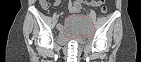
Lymphocele
Seen postoperatively in up to 30% of GYN surgeries with pelvic/para-aortic LND
Believed to arise from surgical transsection or inadequate ligation of draining lymphatics
Lymphatic fluid may accumulate in various pelvic and retroperitoneal compartments
Small lymphoceles typically resorb spontaneously
Large lymphoceles may cause compression symptoms, and may result in abdominal distention, abdominal and pelvic pain, hydronephrosis, bladder dysfunction, constipation, tenesmus, edema of the ipsilateral leg and of the genitalia, and thromboembolism of iliac vessels
Infection may cause fever, chills, and sepsis
Symptomatic lymphoceles are typically managed initially with percutaneous CT-guided drainage, surgical therapy may be required