Medical Physiology/Cellular Physiology
Introduction[edit | edit source]
Chapters[edit | edit source]
- Cell structure and Function
- Membrane Dynamics
- ATP and Energy Production
- DNA and Reproduction
- Gene Expression and Protein Synthesis
- Cell junctions and Tissues

.


Overview[edit | edit source]
All living things are composed of cells which fall basically into two types eukaryotic, or prokaryotic. Bacteria are prokaryotic cells. Typically there is no nucleus, and the intracellular structure lacks organules.
Animals and plants are composed of Eukaryocytic cells, These cells contain a nucleus and are characterized by several different intra-cellular organules. This section deals mainly with animal eukaryocytic cells, although prokaryotic metabolism is touched on because of its importance in antibiotic action.
The thumbnail on the right shows the typical features of a cell as seen under with a light microscope using a standard stain. The chief features are a cell membrane, a granular cytoplasm, and a nucleus with in most cases a darker staining nucleolus.
To see other features usually requires special stains, or an electron microscope.
The chief constituents of the cytoplasm are water, electrolytes, fats, carbohydrates and proteins. Water constitutes about 80% of the cell.
The chief electrolytes are potassium, phosphates sulfates, magnesium and bicarbonates. In smaller concentrations are sodium, calcium and chlorine. Sodium and chlorine are found outside the cell in the interstitial fluid, in the same proportions as sea water, but less concentrated. It is hypothesized that the concentration seen probably represents the concentration of sea water when multicellular animals first evolved from single celled animals.
Fats include the fat soluble phospho-lipids and cholesterol. Phospholipids are of importance because they makeup the membrane of the cell wall, the nuclear membrane, and of the various organules found in the cell.
Carbohydrates include glucose and glycogen. Most cells have some glycogen - about 1%. Muscle cells have about 3%, and the liver up to 7%.
Apart from water, proteins are the next bigest constituent, between 15% and 20%. They fall into two categories, structural proteins and functional proteins. The structural proteins provide the skeleton of the cell, the functional proteins include numerous 'working' polypeptides including enzymes. The cell wall is studded with specialized proteins, indeed the cell wall is about 70% protein. Membranes of organules also have these specialized proteins in their wall. Many of these are concerned with transport of substances in and out of the cell.
In the next section we will look at the makeup and structure of the cell in more detail, and then we will look at how structure is married to function.
Cell Anatomy[edit | edit source]
See Cell structure and Function
Cell Functions and Energy Requirements[edit | edit source]
Cell Membrane Structure[edit | edit source]
Cell Cytoplasm Functions[edit | edit source]
Glycolysis[edit | edit source]
Polypeptide Synthesis[edit | edit source]
Cell Membrane Dynamics[edit | edit source]
Diffusion and Active Transfer[edit | edit source]
Cell Membrane Receptors and Ligands[edit | edit source]
Channels, Pumps & Gates[edit | edit source]
Symports & Antiports[edit | edit source]
Na+/K+ pump[edit | edit source]
Membrane Potential[edit | edit source]
Na/Glucose Cotransporter[edit | edit source]
Ligands and Receptors[edit | edit source]
Pinocytosis[edit | edit source]
Nuclear Function[edit | edit source]
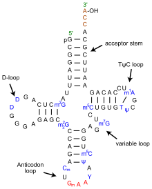
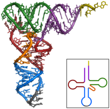
DNA, mRNA & tRNA[edit | edit source]
Wikipedia article on tRNA[1]

S. H. Kim,1 J. L. Sussman,1 F. L. Suddath,2 G. J. Quigley,2 A. McPherson,2 A. H. J. Wang,2 N. C. Seeman,2 and Alexander Rich2
1Department of Biochemistry, Duke University School of Medicine, Durham, North Carolina 27710
2Department of Biology, Massachusetts Institute of Technology, Cambridge, Mass. 02139

Protein Synthesis[edit | edit source]
Mitochondrial Function[edit | edit source]
Citric Acid Cycle[edit | edit source]
Hydrogen Transfer System[edit | edit source]
Centrioles and Cell Division[edit | edit source]
Mitosis[edit | edit source]


Wikipedia article[2]
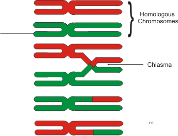
Meiosis[edit | edit source]
Wikipedia article[3]


Primary & Secondary Messengers[edit | edit source]
Tissues[edit | edit source]
Junctions[edit | edit source]


