IB Biology/Cells
Topic 2: Cells[edit | edit source]
2.1 Cell Theory[edit | edit source]
2.1.1 Outline the Cell Theory
- Living cells are composed of one or more cells
- Cells are the smallest unit of life
- All organisms come from pre-existing cells
2.1.2 Discuss the evidence for the cell theory
The cell theory has amassed tremendous credibility through the use of the microscope in the following:
- Robert Hooke- studied cork and found little tiny compartments that he called cells
- Antonie van Leeuwenhoek- observed the first living cells, called them 'animalcules' meaning little animals
- Schleiden- stated that plants are made of 'independent, separate beings' called cells
- Schwann- made a similar statement to Schleiden about animals
- Rudolf Virchow- discovered that all organisms come from pre-existing cells
2.1.3 State that unicellular organisms carry out all of the functions of life
- Metabolism; chemical reactions inside the cell, including cell respiration to release energy
- Response; perceiving and responding to changes in the environment
- Homeostasis; keeping conditions inside the organism within tolerable limits
- Growth; an irreversible increase in size
- Reproduction; producing offspring either sexually or asexually
- Nutrition; obtaining food, to provide energy and the materials needed for growth
- Defense; protection against enemies
Amoeba would be an example of an unicellular organism.
2.1.4 Compare the relative sizes of molecules, cell membrane thickness, viruses, bacteria, organelles and cells, using the appropriate SI unit
nm = nanometer µm = micrometer
- Molecules - 1 nm
- Thickness of membrane - 10 nm
- Viruses - 100 nm
- Bacteria - 1 µm
- Organelles - up to 10 µm
- Most cells - up to 100 µm (three dimensional nature/shape)
2.1.5 Calculate the linear magnification of drawings and the actual size of specimens in images of known magnifications
- :Drawings should show cells and cell ultrastructure.
- Include:
- A scale bar: |------| = 1 µm
- Magnification: ×250
- To calculate magnification:
- Magnification = Measured Size of Diagram ÷ Actual Size of Object
but before this, both magnifications must be in the same measuremente, either in mm, cm etc..
2.1.6 - Explain the importance of the surface area to volume ratio as a factor limiting cell size.
- A cell needs a large surface area in order to carry out metabolic functions (as chemical reactions require a surface). As a cell grows, it needs to carry out more and more reactions. Therefore, since a cell has to maintain a certain surface area to volume ratio, its size is limited.
- The rate of exchange of materials (nutrients/waste) and energy (heat) is a function of its surface area.
- Thus: As a cell grows in size (volume), the distance increases between the cytoplasm at the center of the cell and the cell membrane. The rate of chemical exchange with the surrounding environment may hence become too low to maintain the cell. It is not able to excrete waste quickly enough or take in important minerals.
- Volume of a cell determines requirements while surface area determines supply.
2.1.7 - State that multicelluar organisms show emergent properties
- Emergent properties arise from the interaction of component parts: the whole is greater than the sum of its parts
2.1.8 - Explain how cells in multicellular organisms differentiate to carry out specialized functions by expressing some of their genes but not others.
- During the early development stages of multicellular organisms, cells undergo differentiation, becoming specialized in structure and function. These cells are then organized into tissues and organs. Cells of multicellular eukaryotes express only a small fraction of their genes, allowing them to perform highly specialized functions. Cells, such as those of muscle or nervous tissue, express only a tiny fraction of their genes.
2.1.9 - State that stem cells retain the capacity to divide and have the ability to differentiate along different pathways.
- Unspecialised cells that can become any type of cells.
- Embryo cells are "totipotent", meaning they can become any cells; after divisions, when the zygote became a ball of cells of blastocyst, which is "pluripotent", meaning capable of being almost any type of tissue. Stem cells can also come from umbilical cord of new baby, which are "multipotent", meaning they can be limited number of tissues.
- Stem cells are self-sustaining: they can divide for many times.
- They differentiate into specific tissue based on a chemical signal
2.1.10 - Outline one use of therapeutic stem cells .
- Bone marrow transplants. They only work because what you are actually transplanting is the hematopoetic stem cells in the marrow. And peripheral blood stem cells, as well as cord blood stem cells, can be used in lieu of bone marrow, making being a donor FAR easier today than in decades past.
Random Stuff that Doesn't fit or is old syllabus material
State that a virus is a non-cellular structure consisting of DNA or RNA surrounded by a protein coat.
- Viruses are not cells. They are simple particles consisting of DNA and RNA wrapped in a protein coat. Viruses are not considered alive because they have no metabolism and they require a host to live. Viruses do not carry out all the functions of life, therefore they are not living.
Explain three advantages of using light microscopes.
- Light microscopes
- Display color instead of monochrome (black and white) images.
- Provide a large field of view.
- Facilitate preparation of sample material.
- Allow for the examination of living material and the observation of movement.
- Cheap in comparison to electron microscopes
Outline the advantages of using electron microscopes.
- Electron microscopes:
- Provide images of higher resolution and magnification than light microscopes.
- Resolution refers to the ability to distinguish two objects as separate entities.
- Magnification refers to the ability to increase the size of a viewed object.
- Scanning Electron Microscopes (SEM) provide images of the specimen's surface while Transmission Electron Microscopes (TEM) provide images of a sample's interior. The resolution of an SEM is approximately half that of a TEM.
- May provide a three dimensional view.
Define organelle.
- An organelle is a discrete structure within a cell, and has a specific function. A mitochondrion would be an example of an organelle.
Organelle List:
ribosome In contrast to the other organelles, they are not surrounded by a membrane.
centriole (Unique to animal cells)
chloroplast (Unique to plant cells)
Define tissue, organ and organ system.
- Tissue: An integrated group of cells that share structure and are adapted to perform a similar function.
- Organ: A combination of two or more tissues which function as an integrated unit, performing one or more specific functions.
- Organ system: A group of organs that specialize in a certain function together.
2.2 Prokaryotic Cells[edit | edit source]
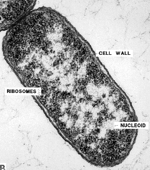
2.2.1 Draw and label a diagram of the ultrastructure of Escherichia (E. coli) as an example of a prokaryote
- The Diagram Should show cell wall, plasma membrane, cytoplasm, pili, flagella, ribosomes and nuceloid (region containing naked DNA)
Two Good Pictures
2.2.2 Annotate the diagram from 2.2.1 with the functions of each of the named structures
- Cell Wall: Maintains the cell's shape and gives protection.
- Plasma Membrane: Regulates the flow of materials (nutrients, waste, oxygen, etc.) into and out of the cell.
- Cytoplasm: Holds and suspends the cell's specialized organelles and enzymes.
- Pili: The function of the pili is attachment to solid surfaces, apparatus for use in transfer of DNA from one cell to another, twitching motility, and cell-cell adhesion.
- Flagella: Flagella are whip like tails that are used to propel the organism forward.
- Ribosome: Protein synthesis.
- Nucleoid: Nucleoid is the area in the cytoplasm where the strands of DNA are present.
2.2.3 Identify structures from 2.2.1 in electron micrographs of E. coli
http://www.ucmp.berkeley.edu/bacteria/bacteriatem.gif http://www.exploratorium.edu/traits/images/ecoli_micro.jpg
2.2.4 State that prokaryotes divide by binary fission.
- Prokaryotes divide by binary fission.
- The process starts with DNA replication, then separation of the two circular strands to either side of the cell. Then cytokenesis occurs: division into two.
- Each new cell has about half of the cytoplasm.
- Growth restores the original size.
Old Stuff not in syllabus or misplaced
State that prokaryotes show a wide range of metabolic activity including fermentation, photosynthesis and nitrogen fixation.
- Prokaryotes demonstrate a range of metabolic activity
- Cyanobacteria (often referred to as blue-green algae although they are not algae) obtain their energy through photosynthesis.
- Bacteria can convert organic substances into other organic substances. (i.e., glucose to lactic acid during anaerobic respiration)
- Some bacteria can fix nitrogen from the air, converting it into ammonia (which is biologically available).
2.3 Eukaryotic Cells[edit | edit source]
2.3.1 Draw and label a diagram of the ultrastructure of a liver cell as an example of an animal cell

- Should include free ribosomes, the rough endoplasmic reticulum (rER), lysosome, Golgi apparatus, mitochondrion, and nucleus.
2.3.2 Annotate the diagram from 2.3.1 with the functions of each of the named structures.
- Ribosomes: Main site of protein synthesis.
- Rough endoplasmic reticulum (rER): Packages the proteins synthesized in the ribosomes.
- Lysosome: Digests macromolecules and contain digestive enzymes.
- Golgi apparatus: Modifies, stores and routes products of the endoplasmic reticulum.
- Mitochondrion: Serves as the site of cellular respiration.
- Nucleus: Contains a cell's genetic material
2.3.4 Compare prokaryotic and eukaryotic cells.
- Differences should include
- Contain naked DNA vs. DNA associated with proteins (DNA wrapped around histones, a protein molecule, creating units called nucleosomes)
- DNA in cytoplasm vs. DNA enclosed in a nuclear envelope - Prokaryotes have naked DNA in cytoplasm, Eukaryotes have a nuclear membrane surrounding it in a nucelus.
- No membrane-enclosed organelles vs. membrane-enclosed organelles (e.g., mitochondria, chloroplasts) - Prokaryotes have no mitchondria, thousands of reactions occur in cytoplasm.
- 70S vs. 80S ribosomes - Prokaryotes = 70S, Eukaryotes = 80S.
- Eukaryotic cells have internal membranes that compartmentalize their functions
- prokaryotic is usually smaller in size, Eukaryotic is larger
- both have cytoplasm
- prokaryotic has no nucleus, Eukaryotic has a membrane-bound nucleus
- prokaryotic has one chromosome / circular, Eukaryotic has two or more chromosomes
- prokaryotic has DNA only, Eukaryotic has DNA with histones to bind together
- prokaryotic has no membrane-bound organelles, E has some membrane-bound organelles
- Eukaryotic has mitochondria, prokaryotic does not
- Eukaryotic has other example of organelle, prokaryotic does not
- both can have a flagellum
- if flagella then E has 9+2 fibrils, prokaryotic does not
- prokaryotic can have plasmids, Eukaryotic does not
- both have ribosomes
- prokaryotic has small ribosomes, Eukaryotic has larger ones
- both have cell membrane
- Eukaryotic has centriole, prokaryotic has no centriole
2.3.5 State three differences between plant and animal cells.
- Only plant cells have:
- Cell walls
- Chloroplasts
- Vacuole - more specificially central vacuole
- Plasmodesmata
2.3.6 Outline two roles of Extracellular components
- The plant wall maintains shape, prevents excess water uptake, and holds the whole plant up against the force of gravity.
- Animal cells secrete glycoproteins that form the extracellular matrix (ECM). This functions in support, adhesion and movement.
Stuff not in syllabus or not relevant or misplaced
- Starch granules for storage of carbohydrates
- Centrioles
- Cholesterol in the plasma membrane
- Glycogen for storage of carbohydrate
State the composition and function of the plant cell wall
- The main component of plant cell walls is cellulose. Cellulose molecules are arranged in bundles called microfibrils. These give the cell wall great tensile strength and allow high pressures to develop inside the cell.
- extra:The cellulose cell wall consists of three layers: middle lamella, primary cell wall, and secondary cell wall. The overall functions the cell wall preforms are: structure, support, protection.
2.4 Membranes[edit | edit source]
2.4.1 Draw and label a diagram to show the structure of membranes.
- Diagram should show the phospholipid bilayer, cholesterol, glycoproteins, and integral and peripheral proteins. Use the term plasma membrane not cell surface membrane for the membrane surrounding the cytoplasm. [1]
![[1]](http://www.proprofs.com/flashcards/upload/a6645014.gif)
2.4.2 Explain how the hydrophobic and hydrophilic properties of phospholipids help to maintain the structure of cell membranes.
- Hydrophilic molecules are attracted to water. Hydrophobic molecules are not attracted to water, but are attracted to each other. The phosphate head is hydrophilic and the two hydrocarbon tails are hydrophobic. In water, phospholipids form double layers with the hydrophilic heads in contact with water on both sides and the hydrophobic tails away from the centre. The attraction between the heads and the surrounding water makes membranes very stable.
In your answer you should:
- — First discuss PHOSPHOLIPID STRUCTURE
- hydrophobic tail/hydrophillic head
- head made from glycerol and phosphate
- tail made from two fatty acids
- —Then discuss the ARRANGEMENT IN MEMBRANE
- form a bilayer
- heads face outside the membrane/ tails face inside the membrane
- there is a hydrophobic interior and a hydrophillic exterior
- —DRAW A DIAGRAM
- —STATE ADDITIONAL INFORMATION
- phospholipids are held together by hydrophobic interactions
- stabilized by interaction of hydrophillic heads with surrounding water
- fluidity helps the membrane to be stable
- the fluidity allows for breaking and remaking of the membranes
- allows for endo/exocytosis
- the hydrophyllic/hydrophobic layers restrict movement
- phospholipids can move about horizontally.
2.4.3 List the functions of membrane proteins.
- hormone binding sites
- enzymes
- electron carriers
- Channels for passive transport
- Pumps for active transport
- cell to cell recognition
- receptors for neurotransmitters
2.4.4 Define diffusion and osmosis.

- Diffusion: is the passive movement of particles from a region of higher concentration to a region of lower concentration, as a result of the random motion of particles.
- Osmosis: the passive movement of water molecules, across a partially permeable membrane, from a region of lower solute concentration to a region of higher solute concentration.
2.4.5 Explain passive transport across membranes by simple diffusion and facilitated diffusion. Mention channels for facilitated diffusion. A molecule or ion that crosses the membrane by moving down a concentration or electrochemical gradient and without expenditure of metabolic energy is said to be transported passively/diffused. All molecules and ions are in constant motion and it is the energy of motion - kinetic energy - that drives passive transport. Transport of uncharged species across a membrane is dictated by differences in concentration of that species across the membrane - that is, by the prevailing concentration gradient. For ions and charged molecules, the electrical potential across the membrane also becomes critically important. Together, gradients in concentration and electric potential across the cell membrane constitute the electrochemical gradient that governs passive transport mechanisms.
- Facilitated diffusion is diffusion that is "facilitated" by proteins that span the membrane and provide an alternative route or bypass. It is similar to simple diffusion in the sense that it does not require expenditure of metabolic energy and transport is again down an electrochemical gradient. Two major groups of integral membrane proteins are involved in facilitated diffusion:
1. Carrier proteins (also known as permeases or transporters) bind a specific type of solute and are thereby induced to undergo a series of conformational changes which has the effect of carrying the solute to the other side of the membrane. The carrier then discharges the solute and, through another conformational change, reorients in the membrane to its original state. Typically, a given carrier will transport only a small group of related molecules.
2. Ion Channels do not really bind the solute, but are like hydrophilic pores through the membrane that open and allow certain types of solutes, usually inorganic ions, to pass through. In general, channels are quite specific for the type of solute they will transport and transport through channels is quite a bit faster than by carrier proteins. Additionally, many channels contain a "gate" which is functions to control the channel's permeability. When the gate is open, the channel transports, and when the gate is closed, the channel is closed. Such gates can be controlled either by voltage across the membrane (voltage-gated channels) or have a binding site for a ligand which, when bound, causes the channels to open (ligand-gated channels). Ion channels allow currents to be carried across the membrane and are thus of particular importance in the physiology of excitable cells like neurons and muscle cells.
- —BROKEN DOWN
- passive transport requires no energy
- molecules move down a concentration gradient
- water moves by osmosis
- moves from higher solute concentration to lower solute concentration
- small uncharged molecules move by diffusion
- charged molecules move by facilitated diffusion
- facilitated diffusion requires protein channels
2.4.6 Explain the role of protein pumps and ATP in active transport across membranes.
- Active transport is the movement of substances across membranes using energy from ATP. Active transport can move substances against a concentration gradient. Protein pumps in the membrane are used for active transport. Each pump only transports particular substances so cells can control what is absorbed and what is expelled.
- goes against concentration gradient
- requires a protein in the cell membrane /pump/carrier protein
- hydrolysis of ATP / ATP → ADP + phosphate;
- involves a conformational change in the pump / protein
2.4.7 Explain how vesicles are used to transport materials within a cell between the rough endoplasmic reticulum, Golgi apparatus, and plasma membrane.
- vesicle is made by pinching off a piece of membrane
- fluidity of membrane allows this
- vesicles can be used to transport material around inside cells
- proteins are transported in vesicles
- from the rough endoplasmic reticulum to the Golgi apparatus
- from the Golgi apparatus to the plasma membrane
- formation of vesicle from plasma membrane allows material to be taken in
- endocytosis/pinocytosis/phagocytosis is absorption of material using a vesicle
- fusion of vesicle with plasma membrane allows material to be secreted / passed out
- exocytosis is secretion of material using a vesicle
2.4.8 Describe how the fluidity of the membrane allows it to change shape, break and re-form during endocytosis and exocytosis
- In endocytosis part of the plasma membrane is pulled inwards. A droplet of fluid becomes enclosed when a vesicle is pinched off. Vesicles can then move through the cytoplasm carrying its contents.
- In exocytosis vesicles fuse with the plasma membrane. The contents of the vesicles are then expelled. The membrane flattens out again.
====2.1 Cell Theory==== (Yet to be fully updated)
2.5.1 Outline the stages in the cell cycle, including interphase (G1, S, G2, mitosis and cytokenisis)
- Interphase: Divided into 3 phases; Gap Phase 1 (Cell grows larger), Synthesis (Genome is replicated), Gap Phase 2 (separates the newly replicated genome).
- Mitosis: Includes four stages--prophase, metaphase, anaphase, and telophase. Spindle fibers attach to the chromosomes and pull sister chromatids apart. It separates two daughter genomes.
- Cytokinesis: Division of the cytoplasm to form two new cells.
2.5.2 State that tumours (cancers) are the result of uncontrolled cell division and that these can occur in any organ or tissue
- Tumors are formed when cell division goes wrong and is no longer controlled. This can happen in any organ or tissue.
2.5.3 State that interphase is an active period in life of a cell when many metabolic reactions occur, including protein synthesis, DNA replication and an increase in the number of mitochondria and/or chloroplasts.\
- Interphase is an active period in the life of a cell during which many metabolic reactions occur such as protein synthesis, DNA replication and an increase in the number of mitochondria and/or chloroplast.
2.5.4 Describe the events that occur in the four phases of mitosis (prophase, metaphase, anaphase and telophase)
- Prophase: The spindle microtubules are extended from each pole to the equator.
- Metaphase: Chromatids move to the equator and the spindle microtubules from each pole attach to each centromere on opposite sides.
- Anaphase: spindle microtubules pull sister chromatids apart making the centromeres to split. This brings the sister chromatids apart, splitting them into chromosomes. Each identical chromosome is pulled to opposite poles.
- Telophase: Spindle microtubules break down, while chromosomes uncoil and therefore are no longer individually visible. The nuclear membrane now reforms. The cell then is divided by cytokinesis to form two daughter cells with identical genetic nuclei.
2.5.5 Explain how mitosis produces two genetically identical nuclei.
- During prophase, chromosomes become visible under a light microscope as they super coil and therefore they get shorter and becomes more bulky. The nuclear envelope disintegrates and the spindle microtubules grow, moves towards each pole, towards the equator. At metaphase the chromatids move to the equator. The sister chromatids are two DNA identical molecules as they were replicated during DNA replication. These sister chromatids are then separated in anaphase as the spindle microtubules attaches to the centromere, it pulls the sister chromatids to opposite poles. Now that sister chromatids are separated, they are known as chromosomes. This means that each pole has the same chromosomes. Finally the microtubules break down, the chromosomes uncoil and the nuclear membrane reforms. The cell then divides into two daughter cells with genetically identical nuclei.
2.5.6 State that growth, embryonic development, tissue repair and asexual reproduction involve mitosis.
- Growth, embryonic development, tissue repair and asexual reproduction involve mitosis.