Sensory Neuroscience: Hearing and speech/Outer & middle ear/tympanic membrane
The eardrum (also tympanic membrane or tympanum), is a thin membrane that separates the external ear from the middle ear. Its function is to transmit sound from the air to the ossicles inside the middle ear. The malleus bone bridges(hammer,anvil) the gap between the eardrum and the other ossicles, and is attached along its long arm on the inner surface. As it is connected to the cardiac nerves of brain.
Structure
[edit | edit source]
The typanic membrane is formed of a double-layer of epithelial cells across it's entire surface. In addition, about 1/3 has another 2 layers of tougher, fibrous epithelial tissue (between the previously-mentioned two layers of cells), to which the long arm of the malleus is attached.
The section with only two layers of cells is pars flaccida, and is flaccid, weak, transparent, and damage to it causes no issues for hearing since it is not involved with transduction. When surgeons perform middle ear surgery, going through pars flaccida is the standard entrance.
The thicker section is pars tensa, and this is the section which transduces the vibrations. Damage to this section of the membrane results in altered hearing.
The umbo (C) is the point of deepest recession of the membrane and is the location of genesis of new epithelial cells, which migrate out towards the pinna as new cells are formed behind them. This migration occurs at about the same rate as the growth of fingernails. In addition, the malleus is attached along the length of its long arm to the inner surface, beginning at the umbo (so, the handle of the malleus is attached at the umbo).
The whole tympanic membrane is about across, with a surface area of (not circular).


-
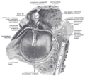 Here, pars tensa is labelled as "tense portion of membrana tympani" and pars flaccida labelled as "flaccid portion of membrana tympani"
 -


