Science: An Elementary Teacher’s Guide/The Human Body: The Skeletal System
Skeletal System
[edit | edit source]
The human skeleton provides the framework and the structural of the human body. It consists of mainly bones that are cushioned by cartilage, even though the bones are made up of cells there is a material that is made up off calcium phosphate that is between the cells that cause bones rigid. Most of the bones are hollow but they are filled with marrow, which is a soft tissue that manufactures red blood cells. The human fetus at first is made up of cartilage for the most part but as the body continues to grow and to mature the cartilage cells are replaced by bone cells. Even though most of the cartilage is turned to bone cells human still have a little of that soft and flexible but tough material in the body. These is located at the end of the long bones it is the padded layer material. Another thing is the human body has discs made from cartilage these provides a cushion between the vertebrae of the spinal column. Fun fact the ears and nose of the human body are also made up of cartilage but a different kind which helps with the form and flexibility.
The Rib Cage
[edit | edit source]
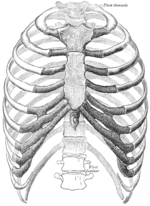
The rib cage, consisting of the sternum (breast bone) and ribs, surrounds the heart, lungs, liver, and spleen. The rib cage contains 24 ribs, symmetrically arranged in 12 matching pairs. The first 7 pairs use the sternum as their anchor via the rib’s individual costal cartilage as an attachment vessel. The first 7 pairs are also called true ribs. Ribs 8 through 12 are deemed false ribs. The remaining 2 pairs of ribs are considered floating ribs as there is no attachment to the sternum at all. True ribs connect directly to the sternum while false ribs are connected to the sternum via additional bone structures, and floating ribs do not connect to the sternum. The sternum is the long and flat bone structure that is actually a plate consisting of 3 separate bones. Consisting of the upper manubrium, the central body, and the lower xiphoid process, the sternum creates an anchor for the ribs meeting centrally at the midline of the body.
The Skull
[edit | edit source]
The human skull or the cranium is an important part of the human skeletal system. The skull is made up of the cheeks, nose, and jaw bone plus other several platelike bones that come together to make up the skull. The human body is made up of over 200 bones but the only one that protects the brain is the skull. When it comes to children the bones are rather loosely due to the fact that they are not done growing up. This way the skull is allowing growth to take place for the child to mature and slowly fuse together the plates to become strong and solidly firm.
The Spinal Column
[edit | edit source]
The human skeletal system is also consisted of the spinal column this helps support the torso and the head which allows them both to bend and turn. The spinal column is made up of a series of thirty three vertebrae. Another major role that the spinal column plays is it provides a protective channel for the spinal cord, each vertebra has a little size donut-hole in the center this helps the spinal cord attach to the base of the brain it goes all the way down to the through the spinal column. Nerves branch out from the spinal column which helps the brain when sending chemical electrical signals through out the whole body.
The Arms and Legs
[edit | edit source]
The arms attach to the pectoral girdle, which makes up the shoulders. The legs attach to the pelvic girdle, which attaches to the lower part of the spinal column and forms the hips. In both the arms and the legs, the half closest to the body has one strong, thick, long bone. The other half of the arm or leg, the part farthest from the body, has two thinner bones. There are 206 bones in the entire human body, so the skeletal structure of the legs and arms are actually quite simple. The hands and feet, though, have many little bones in them.
- Upper Arm
The upper arm has one long bone. This is a strong bone called the humerus. At the shoulder joint, the head of the humerus is shaped like a hemisphere. This shape and the shape of the shoulder joint allows you a large range of movement of the arm. At the elbow joint, it has two protruding bumps called epicondyles, one on the inside of the elbow and one on the outside. When you "hit your funny bone," you are compressing a nerve against the end of the humerus, which gives you the jolt of pain up the arm.
- Forearm
The forearm has two bones in it. These are both long bones, called the radius and the ulna, which both stretch from the elbow to the hand. The radius is shorter and thicker than the ulna though. The radius is on the same side as the thumb and the ulna is on the other side. The range of movement the forearm has is due to the ability of the radius to rotate in the joint it shares with the ulna at the elbow, such as the movement you can see when you lay your forearm flat and flip your hand over and back.
- Thigh
The thigh, like the upper arm, only has one thick, strong long bone. It is called the femur. It has a rounded head sticking out at an angle from the majority of the long bone. This head fits into the hip and allows you a large range of movement of the leg. The end of the femur that fits into the knee joint has two epicondyles, one on each side, just like the humerus in the arm.
- The Kneecap
Compared to the arm, the leg has an extra bone. This is the patella, more commonly known as the kneecap. This small, flat bone is deep in the tendon of a big muscle covering the front of the thigh. It does not interact with the other bones of the leg, but instead helps to arrange the thigh muscle so it works most efficiently.
- Calf
The calf has two long bones in it. The tibia is thicker and longer than the fibula. They are roughly side by side, with the tibia on the big toe side and the fibula on the little toe side. The thin fibula is not part of the knee joint, but rather attaches to the top of the tibia and, at the other end, to the ankle joint.
The Joints
[edit | edit source]A joint is any point where two bones meet. There are three main types of joints; Fibrous (immoveable), Cartilaginous (partially moveable) and the Synovial (freely moveable) joint.
- Fibrous joints
Fibrous (synarthrodial): This type of joint is held together by only a ligament. Examples are where the teeth are held to their bony sockets and at both the radioulnar and tibiofibular joints.
- Cartilaginous
Cartilaginous (synchondroses and sympheses): These joints occur where the connection between the articulating bones is made up of cartilage for example between vertebrae in the spine.
- Synovial Joints
Synovial (diarthrosis): Synovial joints are by far the most common classification of joint within the human body.There are six synovial joints which are: -pivot -hinge -saddle -plane -condyloid -ball & socket joints
