Anatomy and Physiology of Animals/The Gut and Digestion

Objectives
[edit | edit source]After completing this section, you should know:
- what is meant by the terms: ingestion, digestion, absorption, assimilation, egestion, peristalsis and chyme
- the characteristics, advantages and disadvantages of a herbivorous, carnivorous and omnivorous diet
- the 4 main functions of the gut
- the parts of the gut in the order in which the food passes down
The Gut And Digestion
[edit | edit source]Plant cells are made of organic molecules using energy from the sun. This process is called photosynthesis. Animals rely on these ready-made organic molecules to supply them with their food. Some animals (herbivores) eat plants; some (carnivores) eat the herbivores.
Herbivores
[edit | edit source]Herbivores eat plant material. While no animal produces the digestive enzymes to break down the large cellulose molecules in the plant cell walls, micro-organisms' like bacteria, on the other hand, can break them down. Therefore herbivores employ micro-organisms to do the job for them.
There are two types of herbivore:
- The first, ruminants like cattle, sheep and goats, house these bacteria in a special compartment in the enlarged stomach called the rumen.
- The second group has an enlarged large intestine and caecum, called a functional caecum, occupied by cellulose digesting micro-organisms. These non-ruminant herbivores include the horse, rabbit and rat.
Plants are a primary pure and good source of nutrients, however they aren't digested very easily and therefore herbivores have to eat large quantities of food to obtain all they require. Herbivores like cows, horses and rabbits typically spend much of their day feeding. To give the micro-organisms access to the cellulose molecules, the plant cell walls need to be broken down. This is why herbivores have teeth that are adapted to crush and grind. Their guts also tend to be lengthy and the food takes a long time to pass through it.
Eating plants have other advantages. Plants are immobile so herbivores normally have to spend little energy collecting them. This contrasts with another main group of animals - the carnivores that often have to chase their prey.
Carnivores
[edit | edit source]Carnivorous animals like those in the cat and dog families, polar bears, seals, crocodiles and birds of prey catch and eat other animals. They often have to use large amounts of energy finding, stalking, catching and killing their prey. However, they are rewarded by the fact that meat provides a very concentrated source of nutrients. Carnivores in the wild therefore tend to eat distinct meals often with long and irregular intervals between them. Time after feeding is spent digesting and absorbing the food.
The guts of carnivores are usually shorter and less complex than those of herbivores because meat is easier to digest than plant material. Carnivores usually have teeth that are specialised for dealing with flesh, gristle and bone. They have sleek bodies, strong, sharp claws and keen senses of smell, hearing and sight. They are also often cunning, alert and have an aggressive nature.
Omnivores
[edit | edit source]Many animals feed on both animal and vegetable material – they are omnivorous. There are currently two similar definitions of omnivorism:
1. Having the ability to derive energy from plant and animal material.
2. Having characteristics which are optimized for acquiring and eating both plants and animals.
Some animals fit both definitions of omnivorism, including bears, raccoons, dogs, and hedgehogs. Their food is diverse, ranging from plant material to animals they have either killed themselves or scavenged from other carnivores. They are well equipped to hunt and tear flesh (claws, sharp teeth, and a strong, non-rotational jaw hinge), but they also have slightly longer intestines than carnivores, which has been found to facilitate plant digestion. The examples also retain an ability to taste amino acids, making unseasoned flesh palatable to most members of the species.
Classically, humans and chimpanzees are classified as omnivores. However, further research has shown chimpanzees typically consume 95% plant matter (the remaining mass is largely termites), and their teeth, jaw hinge, stomach pH, and intestinal length closely matches herbivores, which many suggest classified them as herbivores. Humans, conversely, have chosen to eat meat for much of the archaeological record, although their teeth, jaw hinge, and stomach pH, and intestinal lengths also closely match other herbivores.
The dispute of human/ chimps classifications is caused by two things. First, there is research that both plant-only and some-animal diets promote health (longevity and freedom from disease) in humans. Second, well-off humans have often chosen to eat meat and dairy products throughout written history, which some argue shows that we prefer meat and dairy by latent instinct.
Per the classical definition, omnivores lack the specialized teeth and guts of carnivores and herbivores but are often highly intelligent and adaptable reflecting their varied diet.
Treatment Of Food
[edit | edit source]Whether an animal eats plants or flesh, the carbohydrates, fats and proteins in the food it eats are generally giant molecules (see chapter 1). These need to be split up into smaller ones before they can pass into the blood and enter the cells to be used for energy or to make new cell constituents.
For example:
- Carbohydrates like cellulose, starch, and glycogen need to be split into glucose and other monosaccharides;
- Proteins need to be split into amino acids;
- Fats or lipids need to be split into fatty acids and glycerol.
The Gut
[edit | edit source]The digestive tract, alimentary canal or gut is a hollow tube stretching from the mouth to the anus. It is the organ system concerned with the treatment of foods.
At the mouth the large food molecules are taken into the gut - this is called ingestion. They must then be broken down into smaller ones by digestive enzymes - digestion, before they can be taken from the gut into the blood stream - absorption. The cells of the body can then use these small molecules - assimilation. The indigestible waste products are eliminated from the body by the act of egestion (see diagram 11.1).
Diagram 11.1 - From ingestion to egestion
The 4 major functions of the gut are:
- 1. Transporting the food;
- 2. Processing the food physically by breaking it up (chewing), mixing, adding fluid etc.
- 3. Processing the food chemically by adding digestive enzymes to split large food molecules into smaller ones.
- 4. Absorbing these small molecules into the blood stream so the body can use them.
The regions of a typical mammals gut (for example a cat or dog) are shown in diagram 11.2.
Diagram 11.2 - A typical mammalian gut
The food that enters the mouth passes to the oesophagus, then to the stomach, small intestine, cecum, large intestine, rectum and finally undigested material exits at the anus. The liver and pancreas produce secretions that aid digestion and the gall bladder stores bile. Herbivores have an appendix which they use for the digestion of cellulose. Carnivores have an appendix but is not of any function anymore due to the fact that their diet is not based on cellulose anymore.
Mouth
[edit | edit source]The mouth takes food into the body. The lips hold the food inside the mouth during chewing and allow the baby animal to suck on its mother’s teat. In elephants the lips (and nose) have developed into the trunk which is the main food collecting tool. Some mammals, e.g. hamsters, have stretchy cheek pouches that they use to carry food or material to make their nests.
The sight or smell of food and its presence in the mouth stimulates the salivary glands to secrete saliva. There are four pairs of these glands in cats and dogs (see diagram 11.3). The fluid they produce moistens and softens the food making it easier to swallow. It also contains the enzyme, salivary amylase, which starts the digestion of starch.
The tongue moves food around the mouth and rolls it into a ball called a bolus for swallowing. Taste buds are located on the tongue and in dogs and cats it is covered with spiny projections used for grooming and lapping. The cow’s tongue is prehensile and wraps around grass to graze it.
Swallowing is a complex reflex involving 25 different muscles. It pushes food into the oesophagus and at the same time a small flap of tissue called the epiglottis closes off the windpipe so food doesn’t enter the trachea and choke the animal (see diagram 11.4).
Diagram 11.3 - Salivary glands
Diagram 11.4 - Section through the head of a dog
Teeth
[edit | edit source]Teeth seize, tear and grind food. They are inserted into sockets in the bone and consist of a crown above the gum and root below. The crown is covered with a layer of enamel, the hardest substance in the body. Below this is the dentine, a softer but tough and shock resistant material. At the centre of the tooth is a space filled with pulp which contains blood vessels and nerves. The tooth is cemented into the socket and in most teeth the tip of the root is quite narrow with a small opening for the blood vessels and nerves (see diagram 11.5).
In teeth that grow continuously, like the incisors of rodents, the opening remains large and these teeth are called open rooted teeth. Mammals have 2 distinct sets of teeth. The first set, the milk teeth, are replaced by the permanent teeth.
Diagram 11.5 - Structure of a tooth
Types Of Teeth
[edit | edit source]All the teeth of fish and reptiles are similar but mammals usually have four different types of teeth.
The incisors are the chisel-shaped ‘biting off’ teeth at the front of the mouth. In rodents and rabbits the incisors never stop growing (open-rooted teeth). They must be worn or ground down continuously by gnawing. They have hard enamel on one surface only so they wear unevenly and maintain their sharp cutting edge.
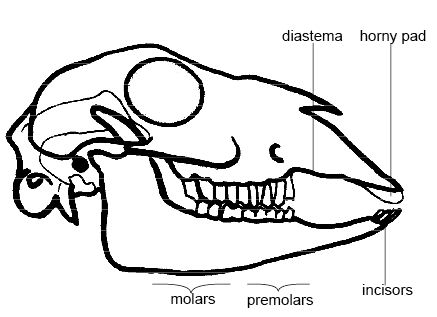
The largest incisors in the animal kingdom are found in elephants, for tusks are actually giant incisors. Sloths have no incisors at all, and sheep have no incisors in the upper jaw (see diagram 11.6). Instead there is a horny pad against which the bottom incisors cut.
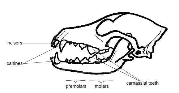
The canines or ‘wolf-teeth’ are long, cone-shaped teeth situated just behind the incisors. They are particularly well developed in the dog and cat families where they are used to hold, stab and kill the prey (see diagram 11.7).
The tusks of boars and walruses are large canines while rodents and herbivores like sheep have no (or reduced) canines. In these animals the space where the canines would normally be is called the diastema. In rodents like the rat and beaver it allows the debris from gnawing to be expelled easily.
The cheek teeth or premolars and molars crush and grind the food. They are particularly well developed in herbivores where they have complex ridges that form broad grinding surfaces (see diagram 11.6). These are created from alternating bands of hard enamel and softer dentine that wear at different rates.
In carnivores the premolars and molars slice against each other like scissors and are called carnassial teeth see diagram 11.7). They are used for shearing flesh and bone.
Dental Formula
[edit | edit source]The numbers of the different kinds of teeth can be expressed in a dental formula. This gives the numbers of incisors, canines, premolars and molars in one half of the mouth. The numbers of these four types of teeth in the left or right half of the upper jaw are written above a horizontal line and the four types of teeth in the right or left half of the lower jaw are written below it.
Thus the dental formula for the sheep is:
- 0.0.3.3
- 3.1.3.3
It indicates that in the upper right (or left) half of the jaw there are no incisors or canines (i.e. there is a diastema), three premolars and three molars. In the lower right (or left) half of the jaw are three incisors, one canine, three premolars and three molars (see diagram 11.6).
Diagram 11.6 - A sheep’s skull
The dental formula for a dog is:
- 3.1.4.2
- 3.1.4.3
The formula indicates that in the right (or left) half of the upper jaw there are three incisors, one canine, four premolars and two molars. In the right (or left) half of the lower jaw there are three incisors, one canine, four premolars and three molars (see diagram 11.7).
Diagram 11.7 - A dog’s skull
Esophagus
[edit | edit source]The Esophagus transports food to the stomach. Food is moved along the esophagus, as it is along the small and large intestines, by contraction of the smooth muscles in the walls that push the food along rather like toothpaste along a tube. This movement is called peristalsis (see diagram 11.8).
Diagram 11.8 - Peristalsis
Stomach
[edit | edit source]The stomach stores and mixes the food. Glands in the wall secrete gastric juice that contains enzymes to digest protein and fats as well as hydrochloric acid to make the contents very acidic. The walls of the stomach are very muscular and churn and mix the food with the gastric juice to form a watery mixture called chyme (pronounced kime). Rings of muscle called sphincters at the entrance and exit to the stomach control the movement of food into and out of it (see diagram 11.9).
Diagram 11.9 - The stomach
Small Intestine
[edit | edit source]Most of the breakdown of the large food molecules and absorption of the smaller molecules take place in the long and narrow small intestine. The total length varies but it is about 6.5 metres in humans, 21 metres in the horse, 40 metres in the ox and over 150 metres in the blue whale.
It is divided into 3 sections: the duodenum (after the stomach), jejunum and ileum. The duodenum receives 3 different secretions:
- 1) Bile from the liver;
- 2) Pancreatic juice from the pancreas and
- 3) Intestinal juice from glands in the intestinal wall.
These complete the digestion of starch, fats and protein. The products of digestion are absorbed into the blood and lymphatic system through the wall of the intestine, which is lined with tiny finger-like projections called villi that increase the surface area for more efficient absorption (see diagram 11.10).
Diagram 11.10 - The wall of the small intestine showing villi
The Rumen
[edit | edit source]In ruminant herbivores like cows, sheep and antelopes the stomach is highly modified to act as a “fermentation vat”. It is divided into four parts. The largest part is called the rumen. In the cow it occupies the entire left half of the abdominal cavity and can hold up to 270 litres. The reticulum is much smaller and has a honeycomb of raised folds on its inner surface. In the camel the reticulum is further modified to store water. The next part is called the omasum with a folded inner surface. Camels have no omasum. The final compartment is called the abomasum. This is the ‘true’ stomach where muscular walls churn the food and gastric juice is secreted (see diagram 11.11).
Diagram 11.11 - The rumen
Ruminants swallow the grass they graze almost without chewing and it passes down the oesophagus to the rumen and reticulum. Here liquid is added and the muscular walls churn the food. These chambers provide the main fermentation vat of the ruminant stomach. Here bacteria and single-celled animals start to act on the cellulose plant cell walls. These organisms break down the cellulose to smaller molecules that are absorbed to provide the cow or sheep with energy. In the process, the gases methane and carbon dioxide are produced. These cause the “burps” you may hear cows and sheep making.
Not only do the micro-organisms break down the cellulose but they also produce the vitamins E, B and K for use by the animal. Their digested bodies provide the ruminant with the majority of its protein requirements.
In the wild grazing is a dangerous activity as it exposes the herbivore to predators. They crop the grass as quickly as possible and then when the animal is in a safer place the food in the rumen can be regurgitated to be chewed at the animal’s leisure. This is ‘chewing the cud’ or rumination. The finely ground food may be returned to the rumen for further work by the microorganisms or, if the particles are small enough, it will pass down a special groove in the wall of the oesophagus straight into the omasum. Here the contents are kneaded and water is absorbed before they pass to the abomasum. The abomasum acts as a “proper” stomach and gastric juice is secreted to digest the protein.
Large Intestine
[edit | edit source]The large intestine consists of the caecum, colon and rectum. The chyme from the small intestine that enters the colon consists mainly of water and undigested material such as cellulose (fibre or roughage). In omnivores like the pig and humans the main function of the colon is absorption of water to give solid faeces. Bacteria in this part of the gut produce vitamins B and K.
The caecum, which forms a dead-end pouch where the small intestine joins the large intestine, is small in pigs and humans and helps water absorption. However, in rabbits, rodents and horses, the caecum is very large and called the functional caecum. It is here that cellulose is digested by micro-organisms. The appendix, a narrow dead end tube at the end of the caecum, is particularly large in primates but seems to have no digestive function.
Functional Caecum
[edit | edit source]The caecum in the rabbit, rat and guinea pig is greatly enlarged to provide a “fermentation vat” for micro-organisms to break down the cellulose plant cell walls. This is called a functional caecum (see diagram 11.12). In the horse both the caecum and the colon are enlarged. As in the rumen, the large cellulose molecules are broken down to smaller molecules that can be absorbed. However, the position of the functional caecum after the main areas of digestion and absorption, means it is potentially less effective than the rumen. This means that the small molecules that are produced there can not be absorbed by the gut but pass out in the faeces. The rabbit and rodents (and foals) solve this problem by eating their own faeces so that they pass through the gut a second time and the products of cellulose digestion can be absorbed in the small intestine. Rabbits produce two kinds of faeces. Softer night-time faeces are eaten directly from the anus and the harder pellets you are probably familiar with, that have passed through the gut twice.
Diagram 11.12 - The gut of a rabbit
The Gut Of Birds
[edit | edit source]Birds’ guts have important differences from mammals’ guts. Most obviously, birds have a beak instead of teeth. Beaks are much lighter than teeth and are an adaptation for flight. Imagine a bird trying to take off and fly with a whole set of teeth in its head! At the base of the oesophagus birds have a bag-like structure called a crop. In many birds the crop stores food before it enters the stomach, while in pigeons and doves glands in the crop secretes a special fluid called crop-milk which parent birds regurgitate to feed their young. The stomach is also modified and consists of two compartments. The first is the true stomach with muscular walls and enzyme secreting glands. The second compartment is the gizzard. In seed eating birds this has very muscular walls and contains pebbles swallowed by the bird to help grind the food. This is the reason why you must always supply a caged bird with grit. In birds of prey like the falcon the walls of the gizzard are much thinner and expand to accommodate large meals (see diagram 11.13).
Diagram 11.13 - The stomach and small intestine of a hen
Digestion
[edit | edit source]During digestion the large food molecules are broken down into smaller molecules by enzymes. The three most important groups of enzymes secreted into the gut are:
- Amylases that split carbohydrates like starch and glycogen into monosaccharides like glucose.
- Proteases that split proteins into amino acids.
- Lipases that split lipids or fats into fatty acids and glycerol.
Glands produce various secretions which mix with the food as it passes along the gut.
These secretions include:
- Saliva secreted into the mouth from several pairs of salivary glands (see diagram 11.3). Saliva consists mainly of water but contains salts, mucous and salivary amylase. The function of saliva is to lubricate food as it is chewed and swallowed and salivary amylase begins the digestion of starch.
- Gastric juice secreted into the stomach from glands in its walls. Gastric juice contains pepsin that breaks down protein and hydrochloric acid to produce the acidic conditions under which this enzyme works best. In baby animals rennin to digest milk is also produced in the stomach.
- Bile produced by the liver. It is stored in the gall bladder and secreted into the duodenum via the bile duct (see diagram 11.14). (Note that the horse, deer, parrot and rat have no gall bladder). Bile is not a digestive enzyme. Its function is to break up large globules of fat into smaller ones so the fat splitting enzymes can gain access the fat molecules.
Diagram 11.14 - The liver, gall bladder and pancreas
Pancreatic juice
[edit | edit source]The pancreas is a gland located near the beginning of the duodenum (see diagram 11.14). In most animals it is large and easily seen but in rodents and rabbits it lies within the membrane linking the loops of the intestine (the mesentery) and is quite difficult to find. Pancreatic juice is produced in the pancreas. It flows into the duodenum and contains amylase for digesting starch, lipase for digesting fats and protease for digesting proteins.
Intestinal juice
[edit | edit source]Intestinal juice is produced by glands in the lining of the small intestine. It contains enzymes for digesting disaccharides and proteins as well as mucus and salts to make the contents of the small intestine more alkaline so the enzymes can work.
Absorption
[edit | edit source]The small molecules produced by digestion are absorbed into the villi of the wall of the small intestine. The tiny finger-like projections of the villi increase the surface area for absorption. Glucose and amino acids pass directly through the wall into the blood stream by diffusion or active transport. Fatty acids and glycerol enter vessels of the lymphatic system (lacteals) that run up the centre of each villus.
The Liver
[edit | edit source]The liver is situated in the abdominal cavity adjacent to the diaphragm (see diagrams 2 and 14). It is the largest single organ of the body and has over 100 known functions. Its most important digestive functions are:
- the production of bile to help the digestion of fats (described above) and
- the control of blood sugar levels
Glucose is absorbed into the capillaries of the villi of the intestine. The blood stream takes it directly to the liver via a blood vessel known as the hepatic portal vessel or vein (see diagram 11.15).
The liver converts this glucose into glycogen which it stores. When glucose levels are low the liver can convert the glycogen back into glucose. It releases this back into the blood to keep the level of glucose constant. The hormone insulin, produced by special cells in the pancreas, controls this process.
Diagram 11.15 - The control of blood glucose by the liver
Other functions of the liver include:
- 3. making vitamin A,
- 4. making the proteins that are found in the blood plasma (albumin, globulin and fibrinogen),
- 5. storing iron,
- 6. removing toxic substances like alcohol and poisons from the blood and converting them to safer substances,
- 7. producing heat to help maintain the temperature of the body.
Diagram 11.16 - Summary of the main functions of the different regions of the gut
Summary
[edit | edit source]- The gut breaks down plant and animal materials into nutrients that can be used by animals’ bodies.
- Plant material is more difficult to break down than animal tissue. The gut of herbivores is therefore longer and more complex than that of carnivores. Herbivores usually have a compartment (the rumen or functional caecum) housing micro-organisms to break down the cellulose wall of plants.
- Chewing by the teeth begins the food processing. There are 4 main types of teeth: incisors, canines, premolars and molars. In dogs and cats the premolars and molars are adapted to slice against each other and are called carnassial teeth.
- Saliva is secreted in the mouth. It lubricates the food for swallowing and contains an enzyme to break down starch.
- Chewed food is swallowed and passes down the oesophagus by waves of contraction of the wall called peristalsis. The food passes to the stomach where it is churned and mixed with acidic gastric juice that begins the digestion of protein.
- The resulting chyme passes down the small intestine where enzymes that digest fats, proteins and carbohydrates are secreted. Bile produced by the liver is also secreted here. It helps in the breakdown of fats. Villi provide the large surface area necessary for the absorption of the products of digestion.
- In the colon and caecum water is absorbed and micro organisms produce some vitamin B and K. In rabbits, horses and rodents the caecum is enlarged as a functional caecum and micro-organisms break down cellulose cell walls to simpler carbohydrates. Waste products exit the body via the rectum and anus.
- The pancreas produces pancreatic juice that contains many of the enzymes secreted into the small intestine.
- In addition to producing bile the liver regulates blood sugar levels by converting glucose absorbed by the villi into glycogen and storing it. The liver also removes toxic substances from the blood, stores iron, makes vitamin A and produces heat.
Worksheet
[edit | edit source]Use the Digestive System Worksheet to help you learn the different parts of the digestive system and their functions.
Test Yourself
[edit | edit source]Then work through the Test Yourself below to see if you have understood and remembered what you learned.
1. Name the four different kinds of teeth
2. Give 2 facts about how the teeth of cats and dogs are adapted for a carnivorous diet:
- 1.
- 2.
3. What does saliva do to the food?
4. What is peristalsis?
5. What happens to the food in the stomach?
6. What is chyme?
7. Where does the chyme go after leaving the stomach?
8. What are villi and what do they do?
9. What happens in the small intestine?
10. Where is the pancreas and what does it do?
11. How does the caecum of rabbits differ from that of cats?
12. How does the liver help control the glucose levels in the blood?
13. Give 2 other functions of the liver:
- 1.
- 2.
Websites
[edit | edit source]- http://www.second-opinions.co.uk/carn_herb_comparison.html Second opinion. A good comparison of the guts of carnivores and herbivores
- http://www.chu.cam.ac.uk/~ALRF/giintro.htm The gastrointestinal system. A good comparison of the guts of carnivores and herbivores with more advanced information than in the previous site.
- http://www.westga.edu/~lkral/peristalsis/index.html Peristalsis animation.
- http://en.wikipedia.org/wiki/Digestion Wikipedia on digestion with links to further information on most aspects. Like most websites this is mainly about human digestion but much is applicable to animals.


