Anatomy and Physiology of Animals/Respiratory System

Objectives
[edit | edit source]After completing this section, you should know:
- why animals need energy and how they make it in cells
- why animals require oxygen and need to get rid of carbon dioxide
- what the term gas exchange means
- the structure of alveoli and how oxygen and carbon dioxide pass across their walls
- how oxygen and carbon dioxide are carried in the blood
- the route air takes in the respiratory system (i.e. the nose, pharynx, larynx, trachea, bronchus,
- bronchioles, alveoli)
- the movements of the ribs and diaphragm to bring about inspiration
- what tidal volume, minute volume and vital capacity are
- how the rate of breathing is controlled and how this helps regulate the acid-base balance of the blood
Overview
[edit | edit source]
Animals require a supply of energy to survive. This energy is needed to build large molecules like proteins and glycogen, make the structures in cells, move chemicals through membranes and around cells, contract muscles, transmit nerve impulses and keep the body warm. Animals get their energy from the large molecules that they eat as food. Glucose is often the energy source but it may also come from other carbohydrates, as well as fats and protein. The energy is made by the biochemical process known as cellular respiration that takes place in the mitochondria inside every living cell.
The overall reaction can be summarised by the word equation given below.
Carbohydrate Food (glucose) + Oxygen = Carbon Dioxide + Water + energy
As you can see from this equation, the cells need to be supplied with oxygen and glucose and the waste product, carbon dioxide, which is poisonous to cells, needs to be removed. The way the digestive system provides the glucose for cellular respiration will be described in Chapter 11 ("The Gut and Digestion"), but here we are only concerned with the two gases, oxygen and carbon dioxide, that are involved in cellular respiration. These gases are carried in the blood to and from the tissues where they are required or produced.
Oxygen enters the body from the air (or water in fish)and carbon dioxide is usually eliminated from the same part of the body. This process is called gas exchange. In fish gas exchange occurs in the gills, in land dwelling vertebrates lungs are the gas exchange organs and frogs use gills when they are tadpoles and lungs, the mouth and the skin when adults.
Mammals (and birds) are active and have relatively high body temperatures so they require large amounts of oxygen to provide sufficient energy through cellular respiration. In order to take in enough oxygen and release all the carbon dioxide produced they need a very large surface area over which gas exchange can take place. The many minute air sacs or alveoli of the lungs provide this. When you look at these under the microscope they appear rather like bunches of grapes covered with a dense network of fine capillaries (see diagram 9.1). A thin layer of water covers the inner surface of each alveolus. There is only a very small distance -just 2 layers of thin cells - between the air in the alveoli and the blood in the capillaries. The gases pass across this gap by diffusion.
Diffusion And Transport Of Oxygen
[edit | edit source]
The air in the alveoli is rich in oxygen while the blood in the capillaries around the alveoli is deoxygenated. This is because the haemoglobin in the red blood cells has released all the oxygen it has been carrying to the cells of the body. Oxygen diffuses from high concentration to low concentration. It therefore crosses the narrow barrier between the alveoli and the capillaries to enter the blood and combine with the haemoglobin in the red blood cells to form oxyhaemoglobin.
The narrow diameter of the capillaries around the alveoli means that the blood flow is slowed down and that the red cells are squeezed against the capillary walls. Both of these factors help the oxygen diffuse into the blood (see diagram 9.2).
When the blood reaches the capillaries of the tissues the oxygen splits from the haemoglobin molecule. It then diffuses into the tissue fluid and then into the cells.
Diffusion And Transport Of Carbon Dioxide
[edit | edit source]Blood entering the lung capillaries is full of carbon dioxide that it has collected from the tissues. Most of the carbon dioxide is dissolved in the plasma either in the form of sodium bicarbonate or carbonic acid. A little is transported by the red blood cells. As the blood enters the lungs the carbon dioxide gas diffuses through the capillary and alveoli walls into the water film and then into the alveoli. Finally it is removed from the lungs during breathing out (see diagram 9.2). (See chapter 8 for more information about how oxygen and carbon dioxide are carried in the blood).
The Air Passages
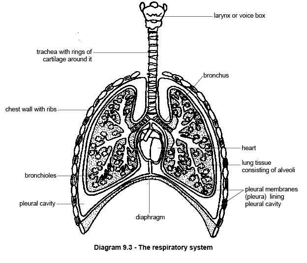
[edit | edit source]When air is breathed in it passes from the nose to the alveoli of the lungs down a series of tubes (see diagram 9.3). After entering the nose the air passes through the nasal cavity, which is lined with a moist membrane that adds warmth and moisture to the air as it passes. The air then flows through the pharynx or throat, a passage that carries both food and air, to the larynx where the voice-box is located. Here the passages for food and air separate again. Food must pass into the oesophagus and the air into the windpipe or trachea. To prevent food entering this, a small flap of tissue called the epiglottis closes the opening during swallowing (see chapter 11). A reflex that inhibits breathing during swallowing also (usually) prevents choking on food.
The trachea is the tube that ducts the air down the throat. Incomplete rings of cartilage in its walls help keep it open even when the neck is bent and head turned. The fact that acrobats and people that tie themselves in knots doing yoga still keep breathing during the most contorted manoeuvres shows how effective this arrangement is. The air passage now divides into the two bronchi that take the air to the right and left lungs before dividing into smaller and smaller bronchioles that spread throughout the lungs to carry air to the alveoli. Smooth muscles in the walls of the bronchi and bronchioles adjust the diameter of the air passages.
The tissue lining the respiratory passages produces mucus and is covered with miniature hairs or cilia. Any dust that is breathed into the respiratory system immediately gets entangled in the mucous and the cilia move it towards the mouth or nose where it can be coughed up or blown out.
The Lungs And The Pleural Cavities
[edit | edit source]
The lungs fill most of the chest or thoracic cavity, which is completely separated from the abdominal cavity by the diaphragm. The lungs and the spaces in which they lie (called the pleural cavities) are covered with membranes called the pleura. There is a thin film of fluid between the two membranes. This lubricates them as they move over each other during breathing movements.
Collapsed Lungs
[edit | edit source]The pleural cavities are completely airtight with no connection with the outside and if they are punctured by accident (a broken rib will often do this), air rushes in and the lung collapses. Separating the two lungs is a region of tissue that contains the oesophagus, trachea, aorta, vena cava and lymph nodes. This is called the mediastinum. In humans and sheep it separates the cavity completely so that puncturing one pleural cavity leads to the collapse of only one lung. In dogs, however, this separation is incomplete so a puncture results in a complete collapse of both lungs.
Breathing
[edit | edit source]
The process of breathing moves air in and out of the lungs. Sometimes this process is called respiration but it is important not to confuse it with the chemical process, cellular respiration, that takes place in the mitochondria of cells. Breathing is brought about by the movement of the diaphragm and the ribs.
Inspiration
[edit | edit source]The diaphragm is a thin sheet of muscle that completely separates the abdominal and thoracic cavities. When at rest it domes up into the thoracic cavity but during breathing in or inspiration it flattens. At the same time special muscles in the chest wall (external intercostal muscles) move the ribs forwards and outwards. These movements of both the diaphragm and the ribs cause the volume of the thorax to increase. Because the pleural cavities are airtight, the lungs expand to fill this increased space and air is drawn down the trachea into the lungs (see diagram 9.4a).
Expiration
[edit | edit source]Expiration or breathing out consists of the opposite movements. The ribs move down and in and the diaphragm resumes its domed shape so the air is expelled (see diagram 9.4b). Expiration is usually passive and no energy is required (unless you are blowing up a balloon).
Lung Volumes
[edit | edit source]
As you sit here reading this just pay attention to your breathing. Notice that your in and out breaths are really quite small and gentle (unless you have just rushed here from somewhere else!). Only a small amount of the total volume that your lungs hold is breathed in and out with each breath. This kind of gentle “at rest” breathing is called tidal breathing and the volume breathed in or out (they should be the same) is the tidal volume (see diagram 9.5). Sometimes people want to measure the volume of air inspired or expired during a minute of this normal breathing. This is called the minute volume. It could be estimated by measuring the volume of one tidal breath and then multiplying that by the number of breaths in a minute. Of course it is possible to take a deep breath and breathe in as far as you can and then expire as far as possible. The volume of the air expired when a maximum expiration follows a maximum inspiration is called the vital capacity (see diagram 9.5).
Composition Of Air
[edit | edit source]The air animals breathe in consists of 21% oxygen and 0.04% carbon dioxide. Expelled air consists of 16% oxygen and 4.4% carbon dioxide. This means that the lungs remove only a quarter of the oxygen contained in the air. This is why it is possible to give someone (or an animal) artificial respiration by blowing expired air into their mouth.
Breathing is usually an unconscious activity that takes place whether you are awake or asleep, although, humans at least, can also control it consciously. Two regions in the hindbrain called the medulla oblongata and pons control the rate of breathing. These are called respiratory centres. They respond to the concentration of carbon dioxide in the blood. When this concentration rises during a bout of activity, for example, nerve impulses are automatically sent to the diaphragm and rib muscles that increase the rate and the depth of breathing. Increasing the rate of breathing also increases the amount of oxygen in the blood to meet the needs of this increased activity.
The Acidity Of The Blood And Breathing
[edit | edit source]The degree of acidity of the blood (the acid-base balance) is critical for normal functioning of cells and the body as a whole. For example, blood that is too acidic or alkaline can seriously affect nerve function causing a coma, muscle spasms, convulsions and even death. Carbon dioxide carried in the blood makes the blood acidic and the higher the concentration of carbon dioxide the more acidic it is. This is obviously dangerous so there are various mechanisms in the body that bring the acid-base balance back within the normal range. Breathing is one of these homeostatic mechanisms. By increasing the rate of breathing the animal increases the amount of dissolved carbon dioxide that is expelled from the blood. This reduces the acidity of the blood.
Breathing In Birds
[edit | edit source]Birds have a unique respiratory system that enables them to respire at the very high rates necessary for flight. The lungs are relatively solid structures that do not change shape and size in the same way as mammalian lungs do. Tubes run through them and connect with a series of air sacs situated in the thoracic and abdominal body cavities and some of the bones. Movements of the ribs and breastbone or sternum expand and compress these air sacs so they act rather like bellows and pump air through the lungs. The evolution of this extremely efficient system of breathing has enabled birds to migrate vast distances and fly at altitudes higher than the summit of Everest.
Summary
[edit | edit source]- Animals need to breathe to supply the cells with oxygen and remove the waste product carbon dioxide.
- The lungs are situated in the pleural cavities of the thorax.
- Gas exchange occurs in the alveoli of the lungs that provide a large surface area. Here oxygen diffuses from the alveoli into the red blood cells in the capillaries that surround the alveoli. Carbon dioxide, at high concentration in the blood, diffuses into the alveoli to be breathed out.
- Inspiration occurs when muscle contraction causes the ribs to move up and out and the diaphragm to flatten. These movements increase the volume of the pleural cavity and draw air down the respiratory system into the lungs.
- The air enters the nasal cavity and passes to the pharynx and larynx where the epiglottis closes the opening to the lungs during swallowing. the air passes down the trachea kept open by rings of cartilage to the bronchi and bronchioles and then to the alveoli.
- Expiration is a passive process requiring no energy as it relies on the relaxation of the muscles and recoil of the elastic tissue of the lungs.
- The rate of breathing is determined by the concentration of carbon dioxide in the blood. As carbon dioxide makes blood acidic, the rate of breathing helps control the acid/base balance of the blood.
- The cells lining the respiratory passages produce mucus which traps dust particles, which are wafted into the nose by cilia.
Worksheet
[edit | edit source]Work through the Respiratory System Worksheet to learn the main structures of the respiratory system and how they contribute to inspiration and gas exchange.
Test Yourself
[edit | edit source]Then use the Test Yourself below to see how much you remember and understand.
1. What is meant by the phrase “gas exchange”?
2. Where does gas exchange take place?
3. What is the process by which oxygen moves from the alveoli into the blood?
4. Why does this process occur?
5. How does the structure of the alveoli make gas exchange efficient?
6. How is oxygen carried in the blood?
7. List the structures that air passes on its way from the nose to the alveoli:
8. What is the function of the mucus and cilia lining the respiratory passages?
9. How do movements of the ribs and diaphragm bring about inspiration? Circle the correct statement below.
- a) The diaphragm domes up into the thorax and ribs move in and down
- b) The diaphragm flattens and ribs move up and out
- c) The diaphragm domes up into the thorax and the ribs move up and out.
- d) The diaphragm flattens and the ribs move in and down
10. What is the function of the epiglottis?
11. What controls the rate of breathing?
Websites
[edit | edit source]A good interactive explanation of breathing and gas exchange in humans with diagrams to label, animations to watch and questions to answer.
Although this is of the human respiratory system there is a good diagram that gives the functions of the various parts as you move your mouse over it. Also an animation of gas exchange and a quiz to test your understanding of it.
- http://en.wikipedia.org/wiki/Lung Wikipedia
Wikipedia on the lungs. Lots of good information on the human respiratory system with all sorts of links if you are interested.
