Three Dimensional Electron Microscopy/Particle Picking
What is Particle Picking[edit | edit source]
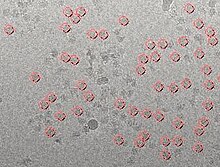
w:Particle picking takes the image obtained from the electron microscopy and its goal it to obtain individual particles from the image. For some particles it works best with certain particle picking programs since many particles have different orientations. Finding individual particles could be done in four ways. The four ways to find individual particles:
- Manual Selection
- Template matching
- Mathematical Function
- Machine Learning
Manual Selection[edit | edit source]

Manual picking is a technique of particle picking that requires the user to manually select the particles from the micrograph. Manual picking is a time consuming technique, which makes it the less preferable over other methods of particle selection. In order to obtain better quality, the user need avoid the particles that are close to the edges, the particles that overlap each other, and the contaminated particles.
Template Matching[edit | edit source]
w:Template matching is a digital technique that is used for object classification. The template matching technique can be used to find particular parts in an image by comparing it to a similar template image. It requires a sample patch image that is used to identify most similar area in the source image [1]. Templates are often used to identify numbers, characters, and any kind of small areas in images. The patch is compared by sliding it on one pixel at a time [2]. Consequently, it calculates an index number that represents how similar the template matches the image in that particular position[1]. The similarity to the template image can be calculated using the correlation coefficient.
Equation to find index number:
Mathematical Function[edit | edit source]
The use of mathematical function helps form templates by using the w:Difference of Gaussian (abbreviated to DoG). DoG picker sorts particle by its size using the radius of a particle and it works along as a reference-free particle picker[3]. The Mathematical function works best with particles that are symmetrical in shape, however not all particles are symmetrical and many are asymmetrical. Unlike manual selection, there are some errors that may occur when using DoG picker. As what was said before some particles are asymmetrical and the DoG picker will have difficulties picking those particles. [3].
Machine Learning[edit | edit source]
Machine learning technique provides automatic particle selection from electron micrograph. Through automatic particle picking, a large number of particles can be selected in a short period of time without the need of extensive manual intervention. The machine learning method can utilize an algorithm to discriminate particles from non-particles [4] The algorithm can be corrected during picking in the training phase to minimize the number of false positives [5] [4]. The training phase has to be semi-supervised to allow the user for algorithm correction to identify the particles selected incorrectly [4]. The method eliminates the need for initial reference volume as it learns the particles of interest from the user.
Reference[edit | edit source]
- ↑ a b Template Matching." Template Matching — OpenCV 2.4.7.0 documentation. N.p., n.d. Web. 22 Nov. 2013
- ↑ Jan Latecki, Longin . "Template Matching." Template Matching. N.p., n.d. Web. 22 Nov. 2013.
- ↑ a b Voss, N.R., C.K. Yoshioka, M. Radermacher, C.S. Potter, and B. Carragher. "DoG Picker and TiltPicker: Software Tools to Facilitate Particle Selection in Single Particle Electron Microscopy." Journal of Structural Biology 166.2 (2009): 205-13. Print.
- ↑ a b c Sorzano C.O.S, Recarte E, Alcorio M, Bibao-Castro J.R., San-Martin C, Marabini R, Carazo J.M., (2009). Automatic particle selection from electron micrograph using machine learning techniques. J Structural Biol. 167 (3), pp.252-260.
- ↑ Zhu Y, Carragher B, Glaeser RM, Fellmann D, Bajaj C, Bern M, Mouche F, de Haas F, Hall RJ, Kriegman DJ, Ludtke SJ, Mallick SP, Penczek PA, Roseman AM, Sigworth FJ, Volkmann N, Potter CS., (2004). Automatic particle selection: results of a comparative study.. J Structural Biol. 145, pp.3-
