Structural Biochemistry/Protein sequence determination techniques/Mass Spectroscopy

Mass spectrometry is an analytical technique used for identifying the mass of a compound based on the mass-to-charge ratio of charged particles. The ratio of charge to mass of the particles is determined by passing them through an applied electric field in a mass spectrometer, which has three main modules: an ion source, a mass analyzer, and a detector. In such procedure, a sample of protein for analysis is placed in the MS instrument. A laser beam is applied to allow the sample to become ionized at the ion source. Positively charged ions of different sizes result and move through the electric field through the analyzer. The lighter ions arrive at the detector first, which triggers a clock to record the time of flight (TOF). This is characteristic of MALDI-TOF mass spectrometry, which can determine the mass of individual components of large protein complexes.
Mass spectrometry is used to analyze the ionized forms of molecules in the gas phase. Mass data is obtained by measuring how fast the ion accelerates through an applied electric field and using Newton's third law, F = ma where F is force, m is mass, and a the acceleration, to calculate the mass since the applied force is known and the acceleration is the experimentally measured value.
Modern techniques for protein analysis include Matrix Assisted Laser Desorption-Ionization (MALDI) and Electrospray Ionization (ESI). Advances to this technique has now made it possible to determine protein masses with an accuracy of one mass unit or less in most cases.
Matrix Assisted Laser Desorption-Ionization (MALDI)[edit | edit source]
The protein or peptide is co-precipitated with an organic compound that absorbs laser light in the matrix. The laser light causes the molecules to expel from the surface and capture electrons as it exits the matrix, leaving the molecules as negatively charged ions. This ionization is very necessary because only ions can be accurately measured. The ionizing laser pulse triggers a clock that measures the time of flight (TOF) for the ions. In time of flight (TOF) analysis, the ions are accelerated through the flight tube in an electric field toward the detector. The lighter ions will arrive first. One of the biggest benefits in using MALDI, as opposed to molecule ionization methods is that MALDI can give molecular fragments, which accurately represent the molecular mass of a protein or peptide. In general weights ranging from a few thousand to several hundred thousand Daltons can be measured.[1]
Electrospray Ionization (ESI)[edit | edit source]

Electrospray Ionization is an ionization technique used for tiny amounts of large molecules such as polymers and proteins, and peptides. The apparatus usually utilizes a hollow metal tube with a sharply pointed end that faces in front of a plate. During the procedure, a sample solution is sprayed, as if from a syringe, from the metal tube, into a strong electric field with the assistance of warm nitrogen for dissolving. The solutions containing proteins or peptides flow through a fine metallic tip at a nonzero electrical potential which releases the solution as electrically charged droplets containing the protein and solvent that evaporates from the droplet, leaving the protein charged at the plate. A great feature of the ESI spectrum is the ability of the ions to carry multiple charges. This technique is often coupled with mass spectrometry for protein analysis.
History of ESI[edit | edit source]
The research on mass spectroscopy started long time ago, but it was not until the 20th century when the electospray ionization technique was developed. The ESI was developed by 2002 Chemistry Nobel Prize winner, Dr. John B Fenn. Together with two other scientists, Tanaka Koichi and Kurt Wuthrich, Dr. Fenn focused the research in the field of mass spectrometry. especially on the ESI technique. Dr. Fenn's research discovery was quickly put into practical use. The ESI brought many benefits during the different usages. For instance, it increased the speed with which complex new pharmaceutical compounds could be evaluated. With this usage, it led to the development of AIDS medications in the mid 1990s.
Properties that make ESI a method of choice for biological applications[edit | edit source]
1. The phage conversion process is "soft"--meaning that it can handle very fragile molecules to be ionized. Furthermore, in some cases, even noncovalent interactions can be put through Mass Spectroscopy.
2. Allows for the analysis of complex mixtures because the eluting fractions can be directly sprayed into the MS
3. Produces natural ions, allowing for the measurement of high-mass biopolymers
[2]

ESI Instruments[edit | edit source]
Quattro II[edit | edit source]
One of the three instruments used for electrospray ionization, Quattro II is the primary instrument used for LR ESI. This instrument is a quadrupole-hexapole-quadrupole mass spectrometer. It has a mass to change rate of 4,000 Da and equipped with an ESI source. During the procedure, samples are inserted into the Quattro II by loop injection or direct infusion through a syringe pump.
LCQ[edit | edit source]
The LCQ instrument is also known as the LCQ Deca XP. This is also an electrospray ionization or ion trap mass spectrometer. It is equipped with the X calibur[check spelling] software that allows acquisition of photodiode array data and mass spectral data. This equipment has a great advantage compared to the other instruments, and it is the capability to perform multiple stages of mass spectrometry. The ability to do this allows an increase in the amount of structural information obtainable for a given molecule. The injection techniques of the LCQ is similar to that of the Quattro, but in slight variations. The LCQ can also be introduced by flow injection using an LC pump or injection valve. Another way is by the LC fitted with a colume[check spelling]. While the Quattro II is the primary instrument for LR ESI, LCQ is the primary instrument for LC/MS and LC/MSMS in ESI.
Q-Tof[edit | edit source]
The Q-Tof is a hybrid quadrupole mass spectrometer with MS/MS capability. Compared to the other two instrument, Q-Tof has a very high resolution, sensitivity, and mass accuracy. With these properties, the Q-Tof is able to assist the mass measurement accuracy for peptides. At the mean time, it can also improved the charge state identification of multiply charged ions and greater differentiation of isobaric species. This instrument can also be equipped with different source. For example, when it is equipped with a nanospray source, it can help to analyze small samples and identify the proteins through semi or complete de novo sequencing. Just like Quattro II and LCQ, Q-Tof can be used by injecting samples through an infusion pump, loop injection, or even an HPLC column. Different from the other two instrument, the Q-Tof is the primary instrument for HR ESI.
New Applications of ESI for Mass Spectrometry[edit | edit source]
Mass spectrometry is an analytical technique used to investigate protein complexes. Recent developments of electrospray ionization mass spectrometry (ESI-MS) have allows characterization of previously unavailable protein complexes, especially those in the gas phase. ESI-MS allows the study of the kinetics of protein complex assembly. Intermediates can be isolated as well as identified using this method. Furthermore, tandem mass spectrometry is used to determine the building blocks that form the global structure of protein complexes. Moreover, ESI-MS is used to identify equilibrium constants by comparing the relative intensity of the complex to the intensity of the subunits. Finally, ion mobility spectrometry mass spectrometry (IMS-MS) is used to analyze macromolecular assemblies. It gives information on the size, shape, mass-to-charge ratio, and number of subunits. This allows the identification of protein rings and protein complex dissociation.
Tandem Mass Spectrometry[edit | edit source]

Individual spectrometer elements or a single mass spectrometer are used in multiple stages of mass analysis separation corresponding to MS steps separated in space or time. In tandem mass spectrometry in space, individual spectrometer elements are separated physically, and these elements can be transmission quadrupole, sectors, or time-of-flight. While in tandem mass spectrometer in time, the separation step is done with multiple steps occurring over a range of time and with the ions trapped in the same place, or space. A quadruple ion trap or FTMS instrument can be applied to such analysis, as it performs analysis on a multiple scale. It is often known as MSn. n refers to the number of steps, hence MS3 refers to a separation composed of three steps.
Approaches for the Analysis of Proteins[edit | edit source]
There are two main ways for the mass spectroscopy analysis of proteins: the bottom-up approach and the top-down approach. In the bottom-up approach, an enzyme, such as trypsin, will be put together with proteins of interest for digestion. The "tryptic peptides" formed as a result of the digestion process are then analyzed by MS and tandem MS. On the other hand with the top-down approach, a protein molecule is analyzed by MS without prior digestion by enzymes. The bottom-up approach is more widely used for several advantages over the top-down approach:
1. Smaller protein ions are more uniform and easy to handle than a whole protein molecule;
2. Masses of smaller protein ions can be determined with higher accuracy;
3. During MS, smaller pieces of proteins are more readily to be reduced into fragments.
However, the one main disadvantage of this approach is the incomplete coverage of the protein molecule. The top-down approach appears to own this advantage while having a lot of other problems, including:
1. The difficulty in handling large protein molecules;
2. Issues about heterogeneity through a whole protein molecule;
3. The complex nature of MS.
Therefore, the top-down analyzes are limited to be applied to only low-throughput single-protein studies. To improve the results of such methods, scientists have come out with an intermediate "middle-down" approach for analyzing proteins that are larger than the smaller protein fragments, such as tryptic pedtides. Although this method is still in development, it is beginning to prove effective, as can be seen in the analysis of modifications on histone tails.[3]
Application of MALDI-TOF MS(mass spectrometry)[edit | edit source]
Usually, the characterization and identification of protein is done by SDS-PAGE (sodium dodecyl sulfate-polyacrylamide gel electrophoresis). However, SDS-PAGE cannot identify small amount of protein samples. MALDI-TOF MS has higher sensitivity and resolution. MALDI-TOF MS can separate protein samples up to 100 kilo Daltons. Therefore, MALDI-TOF MS is used after SDS-PAGE for more accuracy.
First, sample protein is separated by two-dimensional gel. Then, specific cleavage like trypsin cut the proteins. These separated peptides are identified by MALDI-TOF MS. Heavy peptides will slowly move and lighter peptides will move fast due to Newton's second law; F = ma. As peptides move along the tube, the peptides of different accelerations or velocities takes different the amount of time take to reach end of tube. These separated peptides can be analyzed by matching with the computer simulated database.
MALDI-TOF MS also can be used of DNA Hybridization, analyzing microorganism and sugars that bonds with proteins like glycosaminoglycan.
MALDI has many advantageous properties:
1. Robustness
2. High Speed
3. Immunity to contaminants, biochemical buffers, and common additives[4]
Applications of mass spectrometry to lipids and membranes)[edit | edit source]
| ||
||
Lipidomics is an important part of metabalomics is concerned with he detailed depiction and analysis of the structure and function of lipids within a living system. Mass spectrometry has played an extremely crucial and significant role in the determination of the structure of lipids.
Introduction The ever increasing number of completely sequenced genomes available have given biologists the challenge of connecting gene structure to gene function. This drive for understanding the function of genes has spurred scientists to analyze expression levels of the components the make up biological systems like mRNA and proteins. Metabolomics is the study of the endogenously synthesized intermediates called the metabolome and is thought to represent the final result of gene expression. Endogenously synthesized intermediates (metabolomes) are primarily composed of lipids and fats. Studying these lipids can provide information in determining the relationship between the role of lipids and various diseases like cancer, atherosclerosis, and chronic inflammation. In addition, many lipids have cellular membranes and studying their interactions with membrane associated proteins/enzymes can give information on the development of drugs to inhibit these interactions that diseases have.
The first mass spectrometer was constructed by Nobel Laureate Sir J.J Thomson and was used to analyze marsh gas. He observed that the mass to charge rations were 16 to 26, which was identified to be positive ions of methane and acetylene, respectively. Ever since mass spectrometry has been extremely important for the study of proteomics and metabolomics.
Lipid Definition and Classification The categorization of lipids can be wide ranged.It can include organic compounds like fats, oils, waxes, sterol, and tryglycerides, and are insoluble in water but soluble in nonpolar organic compounds. They can also be defined as oily to touch. However, there are many lipids that are not always bound by these definitions. This has led to the official classification of 8 categories of lipids. They are fatty acids, glycerolipids, sphingolipids, sterollipids, prenol lipids, saccharolipids, and polyketides.
Mass Spectrometry in Membrane Protein Analysis[edit | edit source]
Analysis of membrane proteins is challenging because of their hydrophobicity, complex post-translational modifications (PTMs) and relatively low abundance, thus they are not accessible by traditional methods such as X-ray crystallography and NMR spectroscopy. However, with the recent advancements in technology and methodology (ie. better liquid chromatographic performance), mass spectrometry (MS) accelerates membrane protein analysis in a large degree; specifically towards the determination and understanding of the complete plasma membrane (PM) proteome, membrane protein topology, membrane protein-protein interactions and signaling networks originating at the membrane.
MS proteomics to determine the complete membrane proteome[edit | edit source]
The hydrophobicity and low abundance of membrane proteins, and the intricate post-translational modifications has made the analysis of complete membrane proteome very difficult, especially when using traditional approaches, because it is technically challenging to isolate hydrophobic and insoluble proteins. However, with mass spectrometry, coupling with compatible detergents, better instrumentation (such as Multidimensional protein identification technology (MudPIT), which helps to facilitate analysis by separating peptides based on their charge and hydrophobicity), and optimized liquid chromatographic performance, the process of identifying thousands of proteins in one single analysis and get a global overview of all the proteins in the membrane can be done easily and more accurately.
Membrane proteins are generally categorized into three different types:
1. Integral membrane proteins, which is membrane penetrating, is determined to have a few β-barrels (for example, maltoporin) and a majority in α-helical arrangement (for example, the insulin receptor), which can be further divided into four different types based on terminus of the protein and number of times the protein traverses the membrane.
2. Peripheral membrane protein is found to be attached to membrane as in-plane α-helix (such as microtubule-affinity-regulating kinase) or by electrostatic interactions (such as diphtheria toxin).
3. Lipid- anchored proteins is found to be attached to fatty acid, prenyl group or glycophophatidylinositol anchor by covalent bonding (for example the G proteins).
When dealing with detergent-resistant membrane domains and microdomains, mass spectrometry, coupling with multicomplexed fractionation and 1D and 2D gel-based approaches, can also effectively be used to analyze the protein components of the membrane. Studies have shown that these detergent-resistant membranes contain proteins that account for concentrated function.
Protein Topology[edit | edit source]
Instead of using traditional methods such as X-ray crystallography and NMR spectroscopy, mass spectrometry can be used to determine the membrane protein structures, folding and topology at submolecular level, in conjunction with one of the following methods:
1. Hydrogen/deuterium (H/D-MS) exchange
This is based on the hydrogen atom exchange between proteins and the surrounding aqueous solution. And the rate of exchange is ruled by the solvent accessibility through the bilayer of the membrane. New findings using this method are that most of the hydrogen bonding interactions in the protein only moderately stabilized the folded state, and that there are several dynamic regions within the β2 adrenergic receptor (G-protein-coupled receptor).
2. Oxidative or hydroxyl radical probe mass spectrometry
This has the key advantage, over other approaches, in that hydroxyl radicals can be generated directly in solution and react permanently with minimal modification through the incorporation of a limited number of oxygen atoms at reactive residue side chains. See http://en.wikipedia.org/wiki/Protein_footprinting
3. Covalent tagging with regents such as carbodiimide diisopropylcarbodiimide (DiPC-MS)
This is also specific in the residues, Asp and Glu. From this method, it was shown that Glu269 serves an important role in substrate binding of the membrane protein lactose permease.
One main advantage of using MS-based topology approaches is that they are not limited by the type and the size of the membrane proteins.
Membrane protein-protein interactions[edit | edit source]
Mass spectrometry is useful to discover exactly which proteins physically and functionally interact, thus further our understanding of the molecular function of the plasma membrane proteins.
Two approaches are generally used:
1. Isolate membrane protein complexes by antibody purification: Best for identifying multiprotein complexes
With this antibody purification approach, more receptors are identified, such as the α-amino-3-hydroxy-5-methyl-4-isoxazolepropionic acid receptor (AMPAR), γ-aminobutyric acid and kainate receptors, in addition to the previously identified N-methyl-D-aspartate (NMDA) and 5-hydroxytryptamine (5-HT2C) receptors.
2. In vitro binding experiments followed by mass spectrometry: Best for finding direct high-affinity interactions such as ligand and receptor pairs
Signaling Networks Across Membrane[edit | edit source]
Plasma membrane is where all the signaling between cells takes place. Many surface receptors, for example, GPCPs, receptor tyrosine kinases, adhesion signaling molecules and channels, are embedded in the plasma membranes. To further understand membrane signaling, mass spectrometry is used to determine the changes in protein abundance or PTM such as phosphorylation, after treated the cell cultures with growth factor – ligand.
One example of membrane signaling would be the analysis of brassinosteroid (BR) membrane. Arabidopsis thaliana seedling were first treated with BRs. Then the membrane fractions were analyzed using 2D gel and mass spectrometer. The findings showed that membrane-associated kinases of BR transmembrane receptors are responsible for BR membrane signaling. Another example is that the epidermal growth factor signaling of phosphorylated proteins was investigated using MS with the use of stable isotope labeling and affinity capture. From these studies, thousands of proteins were successfully identified.
Quantitative Proteomics[edit | edit source]
Quantitative proteomics is becoming a major part of biological research because it allows scientists to track the dynamic changes of proteins, including post-translational modifications and the formation of protein complexes. This technique often plays a large role in generating new insights and hypothesis for biological processes, which can then be validated by other research approaches. Understanding how proteins change and interact leads to greater understanding in cellular processes and disease progression. For example, it is known that histone regulates gene activation and DNA repair by undergoing methylation, acetylation, and other modifications that changes its interaction with DNA and nuclear proteins. Studying the structural changes of histone during transcription would thus be helpful in learning how to deactivate undesirable genes and amplify beneficial genes. Another heavily targeted area of study is the comparison of protein expression, modifications, and interactions between the diseased state proteins versus that of the normal condition. Understanding these differences can offer valuable insights into the mechanics of disease progression and provide guidance for structure-based drug design. Although tracking protein dynamics is difficult due to the complexity of the biochemical pathways and the drastic changes during different cellular states, the use of mass spectroscopy has greatly reduced these challenges.
Labelling Techniques[edit | edit source]
Before peptide tagging was used to study protein dynamics, quantitative proteomics relied on two-dimensional polyacrylamide gel electrophoresis (2D-PAGE). After running 2D-PAGE, the spot intensities from two or more samples were compared and a mass spectroscopic analysis was used to identify and quantify the protein. Modern quantitative proteomics relies on isotope labelling and mass spectrometry. These methods include:
.jpg)
Isotope-Coded Affinity Tag (ICAT)[edit | edit source]
The protein of interest is purified from different cell states and immersed in labelling reagents that has a biotin tag and a linker region with either heavy or light isotopes. These reagents bind to cysteine-containing peptides, creating a labelled peptide. The proteins are then combined and digested, forming labeled and unlabelled peptide fragments. Combining the digestion process reduces variation in the procedure so that experimental error can be minimized. The labeled peptides are first selected through affinity chromatography based on the presence of the biotin marker, then analyzed with mass spectrometer. Proteins labeled with the light isotope show a lower mass to charge ratio. The relative abundance of the proteins can be determined by the ratio of the peak intensities.
Stable Isotope Labeling with Amino Acids in Cell Culture (SILAC)[edit | edit source]

Stable isotope labeling with amino acids in cell culture (SILAC) is a method of in vivo labeling of proteins for mass spectrometry analysis. The cell culture is grown in a “light” medium and a “heavy” medium. These mediums are given identical conditions except for one particular amino acid that contains a heavy isotope for the “heavy” medium and a light isotope for the “light” medium. Typical isotopes chosen to make the “heavy” medium include 2H, 13C, and 15N. The cells incorporate the heavy or light isotope amino acids naturally during metabolism. After a few generations, every instance of that amino acid will be replaced by the type of amino acid provided in the medium. The proteins grown in both the mediums are then extracted from the cells, purified, and combined together. The proteins are digested with a protease, separated by high performance liquid chromatography, and analyzed by mass spectrometry. As in ICAT, the relative abundance of the proteins can be determined by the ratio of the peak intensities.
Isobaric Labeling[edit | edit source]
Isobaric labeling is a technique that attaches same mass chemical groups to peptides and then using tandem mass spectrometry to determine the relative abundance of the isobaric tag, which represents the abundance of the respectively tagged peptide. Each isobaric tag is composed of a reporter group and a balance group. A peptide-reactive group is attached to the tag to increase peptide affinity to the tag. In each experiment, many isobaric tags can be used to label proteins. All the reporter groups are made to have different masses by varying the amount of 13C, 15N, 18O in the reporter molecule. The balance group is a carbonyl group that is also of varying masses. The overall mass of the reporter group and the balance group are constant for all of the labels. This is useful as an internal standard for mass spectrometry because all labeled proteins should appear in one peak since all the masses are the same. There are two types of isobaric labeling, both utilizing the method as described above:
- iTRAQ (isobaric tag for relative and absolute quantification)
- TMT (Tandem mass tag)
Hydrogen Exchange Mass Spectrometry[edit | edit source]
Hydrogen exchange mass spectrometry is a contemporary method to study protein dynamics, protein-solvent interactions, and protein complex interactions. This technique takes advantage of the exchanging behavior of amide hydrogen to label the protein with deuterium, which can be detected through mass spectrometry or NMR analysis. This method generates information for protein dynamics by describing the solvent accessibility and level of disorder for the various parts of the protein.
Hydrogen-Deuterium Exchange Reaction[edit | edit source]
In solution, the hydrogen covalently bonded to the nitrogen of the peptide (called amide hydrogen) exchanges proton with the solvent. By replacing H2O solvent with D2O, the amide hydrogen can be incorporated into the peptide during the hydrogen exchange process. This is usually accomplished by diluting the protein in H2O buffer with the analog D2O buffer tenfold, thus surrounding the protein with mostly D2O and lessening its contact with H2O. Hydrogen exchange reaction is significantly affected by temperature and pH. Since the exchange process can be acid or base catalyzed, pH must be monitored carefully during the D2O incubation. To study proteins at the natural state, exchange condition is set at pH 7-8 to mirror the physiological environment of the proteins. At pH 2.6, the exchange reaction occurs the slowest. A standard exchange experiment involves immersing the protein for a set amount of time in the D2O buffer, then rapidly shifting the pH of the solution to 2.6, thus slowing the exchange reaction, a process called quenching. A set of exchanging experiments consist of similar exchanging conditions but each experiment is given varying reaction times, commonly ranging from 10 seconds to 3000 seconds or longer. Exchange reactions occur extremely fast at room temperature. To more accurately characterize the deuteration levels among the proteins samples at each of the time points, exchanges are performed at cold temperatures (around 4oC) to decrease the rate of exchange.
Deuterium Detection by Mass Spectrometry[edit | edit source]
To analyze the deuterated proteins with mass spectrometry, the proteins are denatured and digested to form peptides. The peptides are separated by HPLC and analyzed by the mass spectrometer. Since the deuterium nucleus is heavier than a hydrogen nucleus because it contains a neutron as well as a proton, the deuterated peptide has a higher mass compared to the non-deuterated peptide. Based on this mass difference, mass spectrometer can accurately distinguish among the peptides with different deuteration levels. The rate of deuteration for each peptide can also be tracked by measuring the masses of identical peptides that are immersed in D2O for different time points.
Relation to Protein Structure[edit | edit source]
Two factors govern exchange behavior for each peptide: solvent accessibility and hydrogen bonding. Peptides buried in the interior of the protein or surrounded by hydrophobic surfaces show less exchange behavior due to the lack of contact with the D2O solvent. Peptides that form the surface of the protein exchange rapidly and completely because they are in constant contact with D2O. Hydrogen bonding also determines exchange behavior because the hydrogen participating in bonding cannot spontaneously exchange without breaking the bond. Thus great rates of exchange can be attributed to lack of bonding or disorganization. Flexible parts of the protein that are not structured can be characterized by the incorporation of many deuterons. Hydrogen exchange mass spectrometry is also used to study the dynamics of protein complexes to determine which areas of the protein changes in flexibility or solvent accessibility upon binding. This can help identify the areas of the protein that are pertinent to protein-protein interaction and the areas that are dramatically affected by the interaction.
Mass analyzer designs[edit | edit source]
Fourier-transform Mass Spectrometry[edit | edit source]
Fourier-transform mass spectrometry is a type mass spectrometry that takes advantage of ion-cyclotron resonance to select and detect ions. Fourier-transform MS was invented by Alan G. Marshall and Melvin B. Comisarow at the University of British Columbia in 1974. Fourier-transform MS, also known as Fourier transform ion cyclotron resonance MS has high resolution; therefore, it is used as determining the composition of molecules based on accurate mass. It is also capable of studying large macromolecules such as proteins with multiple charges due to its high resolution.

Ion-trap Mass Spectrometry[edit | edit source]
Ion-trap mass spectrometry exists in both linear and 3-D varieties. IT MS was invented by Wolfgang Paul. It uses constant DC and radio frequency oscillating AC electric fields to trap ions in a small volume. This 3-D trap consists of a ring electrode separating two hemispherical electrodes. A mass spectrum is obtained by changing the electrode voltages to eject the ions from the trap. Such technique gives its compact size and the ability to trap and accumulate ions to increase the signal-to-noise ratio of a measurement.

Magnetic-Sector Mass Spectrometry[edit | edit source]
Magnetic-sector mass spectrometry uses a static electric or magnetic sector to affect the path and/or velocity or the charged particles. There are two main types of magnetic-sector MS, single focusing analyzers and double focusing analyzers.

Ion Mobility Mass Spectrometry[edit | edit source]
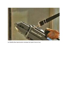
Quantum Sniffer(the figure shown on the right) is an ion mobility mass spectrometry developed by Implant Science Corporation. It is a photonic, non radioactive ionization to detect nano particles in an aqueous solution. The molecular weight, shape, and size of the ionized particles would impact the ion mobility as it pass through drift region and reach the detecting point. The detection of the samples are collected by the vortex, ionized photonically, and analyzed via ion mobility spectrometery (IMS). The samples are collected through a vacuum space, then it is pump into the ion source with electrical current in the ion-molecule reaction region. Then, the ion molecule is then further analyzed based on its drift time, the time for it to go from one chamber to another based on its molecular weight. At the end of the result, computer was able to analyzed a resultant graph of detecting particles.
Advances in Mass Spectrometry[edit | edit source]
The emergence of advances in studying of structural genomics as well as proteomics can yield more information on soluble proteins and their complexes.
An example of a new type of mass spectrometry technique is called laser induced liquid bead ion desorption (LILBID). This technique allows can detect membrane proteins by generatingseries of microdroplets of proteins that then get radiated using a laser into an IR range. At lower intensities, complete complexes can be identified whereas at higher intensities, individual subunits can be detected. This new technique can also be thought of as a combination of another type of mass spectrometry often used, MALDI and electrospray. Despite the idea that LILBID could be a combination of these techniques, there are some key similarities and differences between LILBID itself and ES techniques. Both LILBID and Es are able to explain the stoichiometry of intact membrane protein complexes; however ES has the ability to determine small molecule binding that occurs directly within the complexes. In addition, mass spectrometry studies using this ES technique shows that micelles can indeed exist while in the gas phase. Since LILBID has a lower mass resolution compared to ES, it is unable to provide a more precise characterization of the mass differences between the intact complexes.
Further Improvements Needed for Mass Spectroscopy[edit | edit source]
Although MS technologies have been greatly improved in the last two decades, further improvements are needed for the analysis of protein molecules:
1. Sensitivity of the instrument should be improved in order to analyze smaller number of cells or samples, which allows one to look at corresponding cellular components with their specialized functions;
2. Improved methods are needed to measure low abundance components within protein molecules despite the existence of higher abundance molecules;
3. Higher instrumental speed is needed to allow for deeper and more routine protein analyzes;
4. More robust instruments should be developed to enhance the usage of MS, since some biologists are not quite familiar with these instruments;
5. Improved techniques should be devised to prepare "frozen" samples that are easier to be analyzed.[7]
References[edit | edit source]
- ↑ Template:Citebook
- ↑ Chait, Brian T. "Mass Spectroscopy in the Postgenomic Era." Laboratory for Mass Spectroscopy and Gaseous Ion Chemistry, The Rockefeller University, New York, NY 10021; email chait@rockefeller.edu.
- ↑ Annu. Rev. Biochem. 2011. 80:239-46 The Annual Review of Biochemistry is online at biochem.annualreviews.org
- ↑ Chait, Brian T. "Mass Spectrometry in the Postgenomic Era." Laboratory for Mass Spectroscopy and Gaseous Ion Chemistry, The Rockefuller University, New York, NY 10021; email: chait@rockefeller.edu.
- ↑ Richard Harkewicz and edward A.Dennis(5 April 2011). [1]. "AnnualReview", p. 2-6.
- ↑ Savas, Jeffery, Bejamin Stein, Christine Wu, and John Yates. "Mass Spectrometry Accelerates Membrane Protein Analysis." Trends in Biochemical Sciences. 36.7 (2011): 388-396. <http://dx.doi.org/10.1016/j.tibs.2011.04.005>.
- ↑ Annu. Rev. Biochem. 2011. 80:239-46 The Annual Review of Biochemistry is online at biochem.annualreviews.org
Nelson P. Barrera and Carol V. Robinson. "Advances in the Mass spectroscopy of membrane proteins: From Individual proteins to intact complexes". http://www.annualreviews.org/doi/full/10.1146/annurev-biochem-062309-093307?url_ver=Z39.88-2003&rfr_id=ori:rid:crossref.org&rfr_dat=cr_pub%3dpubmed