File:Lung epithelium 80294-2.6.jpg
Original file (1,600 × 1,286 pixels, file size: 829 KB, MIME type: image/jpeg)
|
|
This is a file from the Wikimedia Commons |
Summary
| Description |
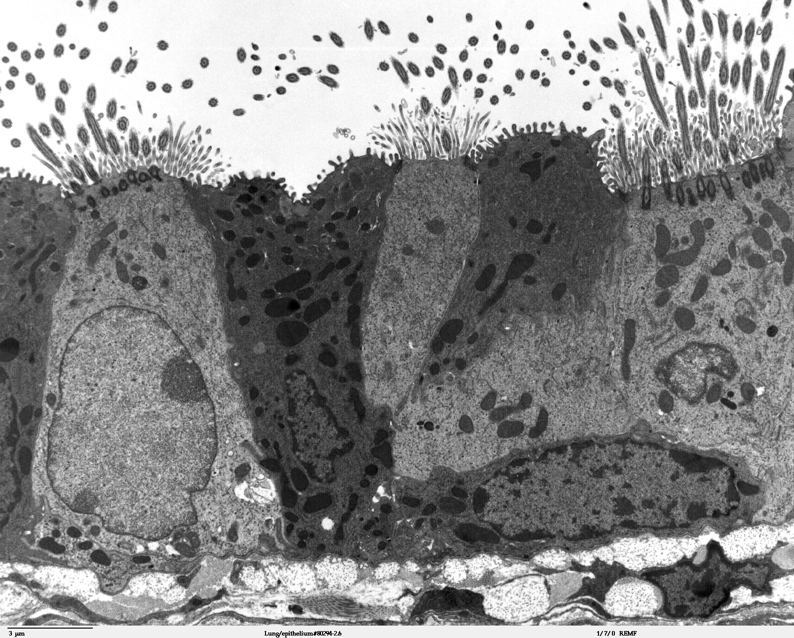
Transmission electron microscope image of a thin section cut through the bronchiolar epithelium of the lung (mouse), which consists of ciliated cells and non-ciliated cells (called Clara cells). Image shows the ciliary microtubules in transverse and oblique section. In the cell apex are the basal bodies that are the anchoring sites for the ciliary axonemes. Note the difference in size and shape between the microvilli and the cilia. JEOL 100CX TEM |
| Source | |
| Author | Louisa Howard, Michael Binder |
| Permission (Reusing this file) |
PD |
Licensing
| This work has been released into the public domain by its author, Louisa Howard and Michael Binder. This applies worldwide. In some countries this may not be legally possible; if so: Louisa Howard and Michael Binder grants anyone the right to use this work for any purpose, without any conditions, unless such conditions are required by law.
|
File history
Click on a date/time to view the file as it appeared at that time.
| Date/Time | Thumbnail | Dimensions | User | Comment | |
|---|---|---|---|---|---|
| current | 19:58, 29 September 2006 | | 1,600 × 1,286 (829 KB) | Patho | {{Information |Description=Transmission electron microscope image of a thin section cut through the '''bronchiolar epithelium of the lung (mouse)''', which consists of ciliated cells and non-ciliated cells (called Clara cells). Image shows the ciliary mic |
File usage
The following page uses this file:
Global file usage
The following other wikis use this file:
- Usage on cs.wikipedia.org
- Usage on de.wikipedia.org
- Usage on de.wikibooks.org
- Usage on es.wikipedia.org
- Usage on lt.wikipedia.org
- Usage on sr.wikipedia.org